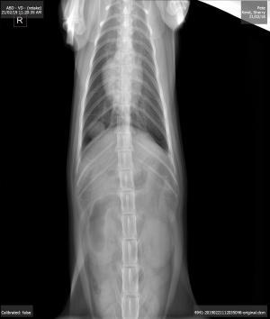
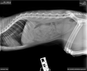
– 1 year old DSH present for three day history of vomiting and not eating
– owner also thought that the patient may have been coughing a so rDVM thought maybe the cat may having been coughing leading to vomiting – history not clear
– bloodwork including FIV/FeLeuk Negative T-39.4C
– x-rays show an approx. 1 cm nodule in the right caudal thorax
– the nodule could not be located on thoracic scan between the ribs even with different positioning and with collapsing the rib cage (the cat was heavily sedated)- only normal lung surface seen
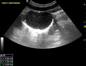
– the nodule could be seen however with a sub-xipohoid approach angling the probe cranially under the rib cage and to the right scanning through the liver and the diaphragm; colour flow not appreciated
– the aorta, cvc and esopahgeal inlet on the left side were all identified and not associated with the mass
– rest of abdominal scan normal except there was some food in the stomach so we suspect the patient has been eating a bit
So what is this? Sampling not possible as lung sliding in and out cranially and liver sliding caudally over the region so risk of laceration. To me it appears to be attached to the thoracic side of the diaphragm and not in the lung. I am not even sure if this is incidental at this time.


Comments
x