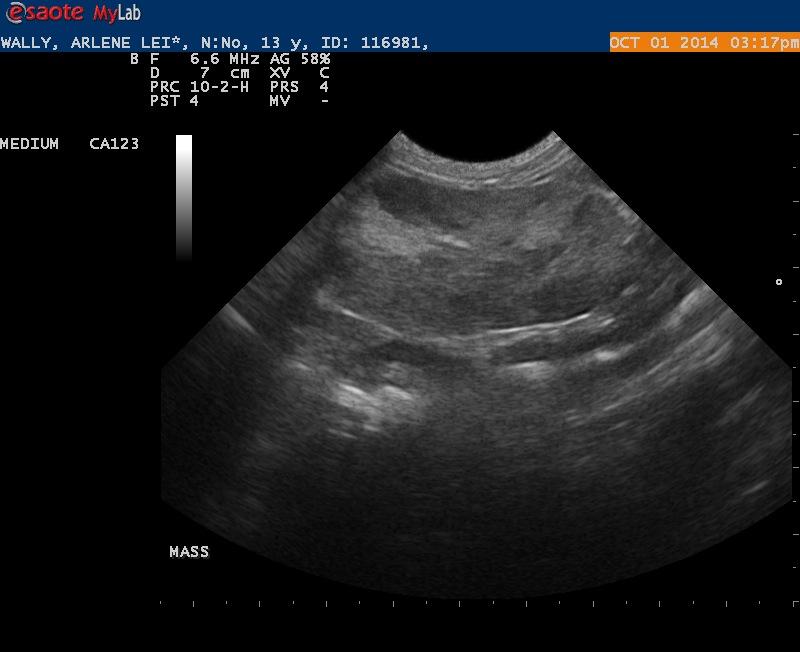
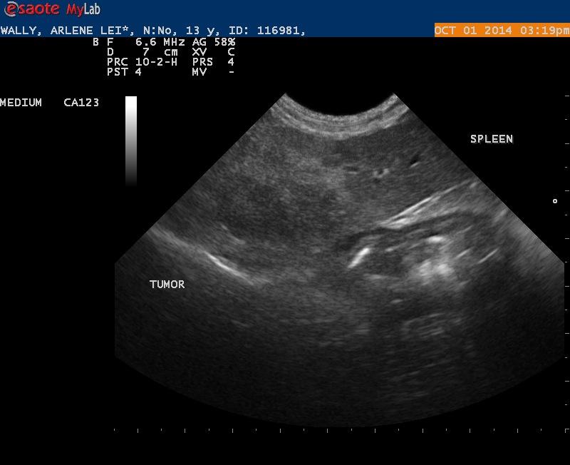
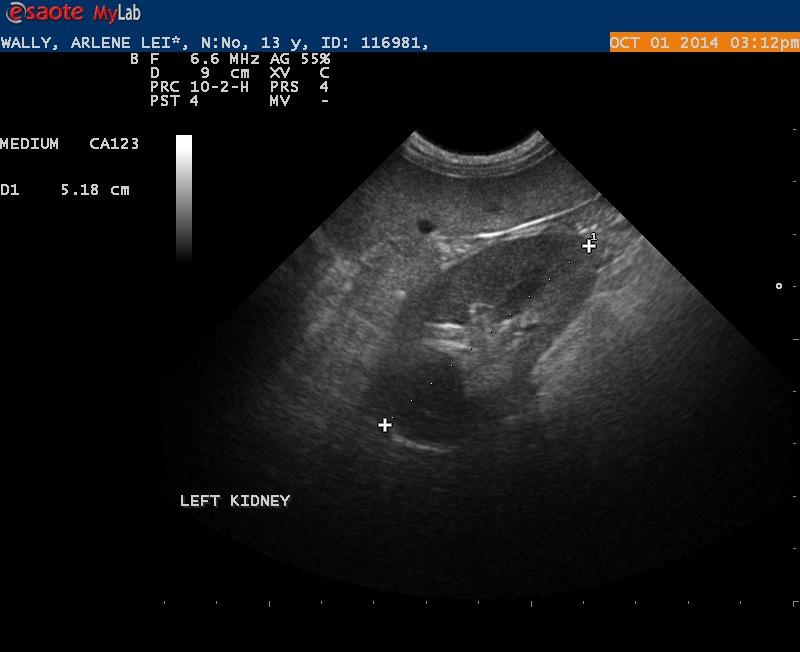
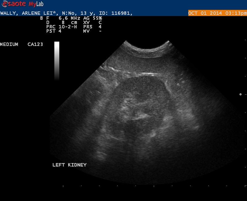
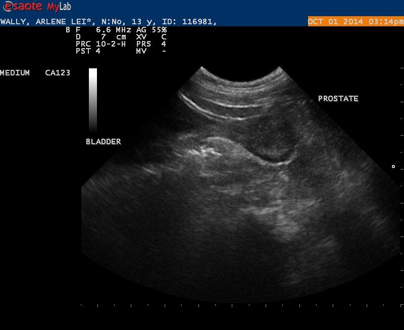
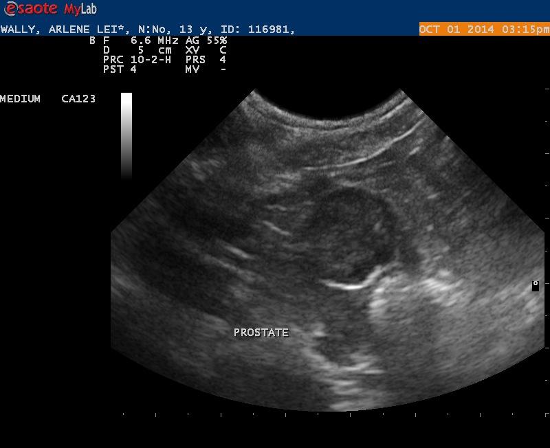
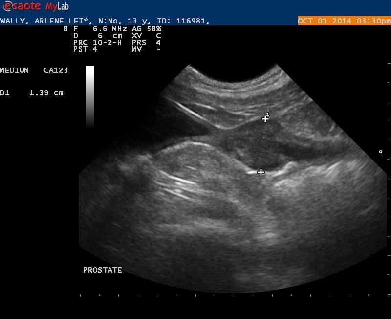
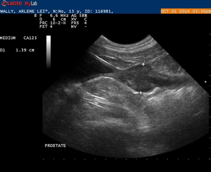
I did an ultrasound on a 13 year old Miniature Schnauzer named Wally. The purpose of the ultrasound was to evaluate Wally for pathology associated with hematuria. On the scan I found a mass that I believe is associated with the spleen. I have a number of questions:
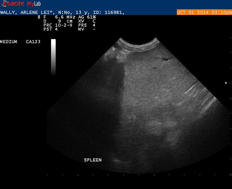
1. Is this mass assoicated with the spleen. I have presented 1 image that I think transitions to healthy spleen
2. Are there nodules in the head of the spleen?
3. There is proteinuria (Prot/Creat of 4.5). Does the renal cortex appear hyperechoic maybe consistent with a glomerulonephritits?
4. Does the prostate look normal?

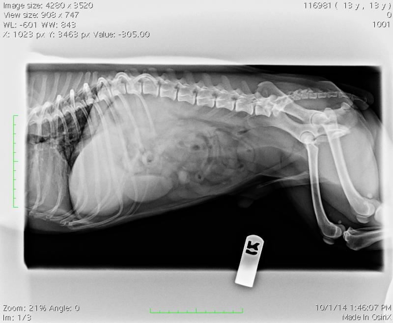
Comments
We had some server delays
We had some server delays so didn’t see these posts getting to them now as we have a 24 hour or less policy on responses:
Yes the mass is surely spleen nice transition image to ensure you can interpret the organ. May be benign hematoma… 50% of these solitary tumors are benign so benefit of doubt but screen the heart for pc effusion or ra mass (15-18% have pc effusion or mass according to our current unpublished study).
Im assuming this is a neutered make and if so the prostate is plump for a MN. Focal mineralization and swollen contour would suggest maybe carcinoma starting but th efact that no peripheral inflammation around the capsule and the capsule is uniform suggests maybe prostatitis but odd for a NM. Needs a needle or traumatic cath. (http://sonopath.com/resources/interventional-procedures) then take out the spleen if benign and heart and chest rads are clean assuming nothing else in the adomen on the sonogram. Kidneys are nsf and may be having secondary proteinuria form the prostate false + or Gn can look like normal kidney (these to me are geriatric normal and looking good for 13) especially with Lyme and other PLN. Nice post:)
Thank EL. Each day is a
Thank EL. Each day is a learning experience.
There was alot of info left out on this case to be brief. The urine had no inflammatory component. The urine Prot/Creat was 4.5. Follow up UA was negative for blood. I still have concern because both urine samples were free catch samples so the hematuria was not iatrogenic.
I previously did an echo on Wally and there was no sign of pericardial effusion and there was no sign of a cardiac mass.
Wally is scheduled for a splenectomy soon- will get an aspriate on the prostate at that time.
Thanks again
cool… i think this is just
cool… i think this is just prominent prostate but will fna fat if thats the case and creamy red if carcinoma typically
cool… i think this is just
cool… i think this is just prominent prostate but will fna fat if thats the case and creamy red if carcinoma typically
Update- I have egg on my
Update- I have egg on my face. This mass was actually in the liver and “butted” up against the spleen. I should have submitted a cine loop. The good news- the mass was removed and hopefully Wally will do well going forward.
Update- I have egg on my
Update- I have egg on my face. This mass was actually in the liver and “butted” up against the spleen. I should have submitted a cine loop. The good news- the mass was removed and hopefully Wally will do well going forward.
Yep well been there done
Yep well been there done that randy and my bad as well I just saw spleen labeled and ran with it without looking foiurther at the infrastructure. If you look at the image attached the arrows indicate thick walled portal veins and the spleen doesnt do this. The capsule technique is still valid here but the capsule is aroundthe liver mass and not splenic mass here. I should have known better to be as definitive on a still because as a rule when reading telemed cases I never ever make a call on a still image so i shouldn’t have here either. Liver and learn. Also the lesion being cranial to the stomach (large arrow) usually is liver unless severe microhepatica is present that brings the spleen cranial to such a position, To scrambled eggs all the way around here 🙁
Thanks EL. I saw the portal
Thanks EL. I saw the portal veins early on and just did not trust what I was seeing.
I really don’t think I will make this mistake again (hopefull). Surgery was still indicated and I am hopeful the histopath will be favorable for Wally.
Thanks EL. I saw the portal
Thanks EL. I saw the portal veins early on and just did not trust what I was seeing.
I really don’t think I will make this mistake again (hopefull). Surgery was still indicated and I am hopeful the histopath will be favorable for Wally.