Rigby is a 10 year 6 month old neutered male Springer Spaniel. He can in febrile and very depressed with a large palpable
mass in the caudal abdomen.
On physical exam he was palpably painful in the caudal abdomen and febrile. Temp was 105.
X-rays revealed a mass in the region of the prostate.
The profile and UA was remarkably normal. No signs of a cystitis. CBC- elevated WBC count- bit over 20,000 with a stress leukogram.
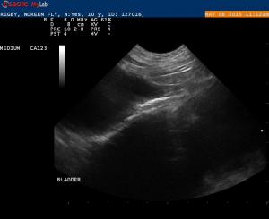
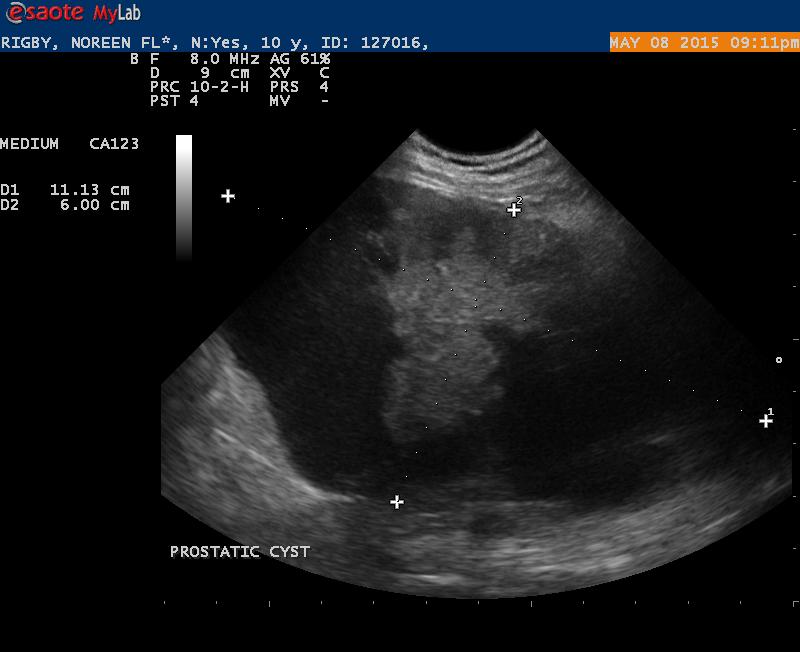
My associate asked me to do an ultrasound on Rigby.
I am posting a couple of .jpg’s and a couple of cines.
I drained 120 ml of bloody fluid off this cyst. I am currently waiting for a culture of the fluid. Cytology did not indicate an abscess. Mostly RBC’s and what I considered to be normal prostatic epitelial cells. I have sent the slides off for cytology. I tried to get my sample from the consolidated area in the middle of the cystic struction. I was unable to drain any more off the cyst. The way it was I was using an 18 guage IV catheter.
My list of differentials at this stage would be a prostatic cyst +/- cyst with a carcinoma.
Any thoughts here. This is the mother of all cysts.
No indication of dysuria/poilakuria.


Comments
Wow thats a doozy… maybe
Wow thats a doozy… maybe neutered with a paraprostatic cyst (Mullerian duct remnant) which often shows no clinical signs because its outside the urinary tract…. I would drain the cyst as much as possible and core bx that parenchyma or address it surgically as a debulk.
Wow thats a doozy… maybe
Wow thats a doozy… maybe neutered with a paraprostatic cyst (Mullerian duct remnant) which often shows no clinical signs because its outside the urinary tract…. I would drain the cyst as much as possible and core bx that parenchyma or address it surgically as a debulk.
Thanks EL.
This dog is sick.
Thanks EL.
This dog is sick. I drained as much as I can. I think I am going to have to
take this dog to surgery.
Thanks EL.
This dog is sick.
Thanks EL.
This dog is sick. I drained as much as I can. I think I am going to have to
take this dog to surgery.
sounds surgical
sounds surgical
sounds surgical
sounds surgical
Update on Rigby. The culture
Update on Rigby. The culture grew a Staph pseudointermedius. Cytology was inconclusive for malignancy.
No chage in the cyst and I am trying to get the owner to take the dog to surgery.
I did a practice echo on this dog. No history of heart disease and NO auscultable mumurs.Below is a report of my findings:
Echo Findings: all values in mm
Contractivity seemed to be normal. No obvious valve changes noted consistent with myomatous change
The L ventricle appeared to be volume overloaded but the L atrium was not enlarged. Trivial tricuspid insufficiency noted on CFI and a small to moderate mitral jet noted on CFI.
M Mode:
IVSd: 9.9 (8.8-9.7)
LVd: 44.4 (32.4-33.9)
PWd: 9.9 (7.1-7.8)
IVSs: 14.2 (13.2-14.2)
LVs: 26.3 (19.9-21.3)
PWs: 16.8 (11.4-12.4)
FS: 40.8
Heart rate: 100
EPSS: 5.8 <7.7
IVSd/LVIDd 0.22 ().22-0.34)
LVIDd/LVPWd 4.5 (3-5)
2D Measurements:
Aorta: 18.7
LA : 26.2
LA/AO 1.4 <1.6
LA Max: 42.4
Doppler: m/sec and mmHg
Aortic Flow 1.3 <2
Aortic Flow PG: 6.9
Pulmonary Artaery Flow: 1.2 < 1.6
Pulmonary Artery Flow PG 4.5
Pulmonary Artery Flow Acc time 87 (52-120)
Pulmonary Artery Ejection Time 215
PA Acc time/Ejection time 0.4 > 0.32
Tricuspid Regurgitation: 0.8 < 2.5
Tricuspid Regurgitation PG: 2.5
Mitral Regurgitation: 5.9
Mitral Regurgitation PG: 140.7
Mitral Valve E: 0.6 (.52-.81)
Echocardiographic findings;
1. Mild eccentric hypertrophy with a moderate volume overload of the L ventricle. Normal L atrial size.
2. Mitral insufficiency without obvious changes occuring in the mitral valve leaflets
3. Trivial tricuspid insufficiency with no indicaton of pulmonary hypertension at this time
4. Stage B2 Chronic Valvular Heart disease
I checked his blood pressure with the doppler and it came out to be about 140-142 mmHg. Good confirmation that my
measurement of the mitral jet was valid.
Question:
1. How often do you see a mitral insufficiency of this degree without hearing a mumur?
2. Could the changes seen be related to a myocarditis and the recent high fever?
Hi Randy
As for your first
Hi Randy
As for your first question, from personal experience (which may not account for much), while practicing my echos in the past (scanning relatives pets, staff pets and anyone else who would let me) I was surprised at how many times I detected MR in the mild-moderate range in which I could not hear a murmur. Some of them eventually went on to develop a murmur and at least one progressed from no murmur with MR, to a murmur with MR and then to a loud murmur, severe MR and CHF within a couple of years. My own Jack Russel (aka the “guinea pig”) has mild-moderate MR, mild AI, mild TR and trace PI with no audible murmur. In one CVD case I sent to a cardiologist, she suggested that a murmur may be louder if the jet is quite eccentric toward the left atrial wall.
Thanks for the reply. I
Thanks for the reply. I believe this murmur is eccentric toward the left atrial wall.
I knew the regurgitation was there but I could not believe it was not audible.
I wonder how many others we are missing.
Thank you Jacqueline
Hi Randy
As for your first
Hi Randy
As for your first question, from personal experience (which may not account for much), while practicing my echos in the past (scanning relatives pets, staff pets and anyone else who would let me) I was surprised at how many times I detected MR in the mild-moderate range in which I could not hear a murmur. Some of them eventually went on to develop a murmur and at least one progressed from no murmur with MR, to a murmur with MR and then to a loud murmur, severe MR and CHF within a couple of years. My own Jack Russel (aka the “guinea pig”) has mild-moderate MR, mild AI, mild TR and trace PI with no audible murmur. In one CVD case I sent to a cardiologist, she suggested that a murmur may be louder if the jet is quite eccentric toward the left atrial wall.
Thanks for the reply. I
Thanks for the reply. I believe this murmur is eccentric toward the left atrial wall.
I knew the regurgitation was there but I could not believe it was not audible.
I wonder how many others we are missing.
Thank you Jacqueline