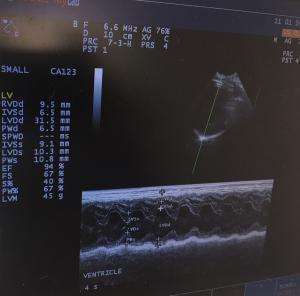
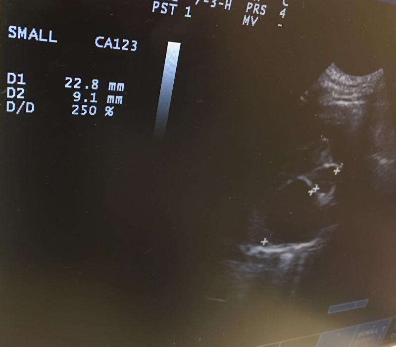
This little Chihuahua is 9 years old and has a grade 4/6 HSM over the mitral valve.
Came in to evaluate a cough. Very limited budget. I just did an M mode of th L ventricle and 2D LA/AO.
In this case there is chamber dilation of the L ventricle with no indication of myocardial hypertrophy- yet the L atrium is grossly dilated.
Here is my questions. Why do some dogs just not respond appropriately to the increased pressure. I can understand an acute situation like a chorde tear- but this is not all that common. Is there an issue at the microscopic level with the myofibrils. This L atrium is not just a little dilated. There does seem to be an appropriate increase in the FS which you would expect. Without the compensatory hypertrophy would you expect a more rapid development of CHF?
Sorry about the quality of the .jpg. I ususally take a picture of my echo’s to work on at home. Any insight would be apprciated.


Comments
May be on the back end of
May be on the back end of compensation… in other words already hypertrophied and then tend to thin out before myocardiual failure occurs with progressive volume overload. Still compensating well now regarding contractility but watch for an eventual drop in fs%.
Hi!
Just to clarify since
Hi!
Just to clarify since this is an interesting topic:
Mitral valve disease does not lead to increased systolic pressures but to increased filling (diastolic) pressures which causes eccentric hypertrophy.
Eccentric hypertrophy means that the heart weight increases without thickening of the myocardium. So, if there is chamber dilation, there is always eccentric hypertrophy present. There is one exception: large PDA: In this case the chamber dilates but the wall thickness usually increase a bit due to the fact that a higher blood volume has to leave the ventricle through the same orifice (aortic valve) – so, the ventricle has to generate more force, which leads to concentric hypertrophy (increase of heart weight due to thickening of the myocardium) along with eccentric hypertrophy.
Thanks for posting
Peter
Thank you Peter. Your
Thank you Peter. Your explanation makes sense. I always thought there had to be wall thickening and chamber dilation to be called Eccentric hypertrophy
I also found the following quick explanation on the internet. You will have to click on it to enlarge.
So what parameters can we rely on when doing an echo to give us a clue that the myocardium is failing. I can think of increased EPSS and a FS that is not compensating properly anymore.