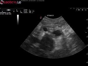
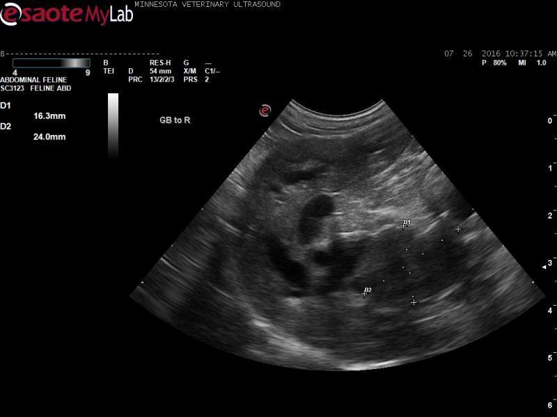
- 4 year old DSH presented for vomiting and possible plastic bag ingestion.
- Chemistry profile showed an ALT>1000 and Total bilirubin=3.8.
- Abdominal ultrasound shows dilated intrahepatic bile ducts, turgid gallbladder with a thickened wall, dilated and tortuous cystic duct, and a 2.5cm hypoechoic mass obstructing the extrahepatic biliary tract. No GI foreign bodies are seen. Urinary bladder calculi are present.
- My primary differential diagnosis for this cat was intially pancreatitis due to the patient’s age. However, upon review, in some clips the mass appears intraluminal (in the bile duct). My complete differential diagnoses list includes pancreatitis, pancreatic neoplasia, bile duct neoplasia (carcinoma) and possibly intraluminal sludge.
- I recommended referral for exploratory surgery with possilbe cholecystectomy or cholecystoduodenostomy.
- Any other thoughts on the pathology of this mass?


Comments
Yes there is a very distinct
Yes there is a very distinct hypoechoic mass obstructing the cbd either panc or cbd origin. Carcinoma usually does this you can fna it and the hepatic parenchyma to check for mets or go in sx to rediect the cbd if no mets in liver but mets are often isoechoic on these and are missed on us. technically possible for panc necrosis here but I doubt it. FNA to be sure. See attached image of your first video.
Hese’s a helpful search on the subject
http://sonopath.com/members/case-studies/search?text=biliary+carcinoma&species=All
Thanks Eric. This cat was
Thanks Eric. This cat was referred to the U where they did a preoperative CT scan. At surgery, the surgeon discovered liver mets (must not have been seen on CT either). The surgeon recommended euthanasia. Unfortunately, histopathology was not performed.
The surgery resident told the client that patients don’t do well after cholecystoduodenostomy (high peri- and post-operative mortality). What is your experience with this procedure with your surgeons out East?
Hmm well im goign to be
Hmm well im goign to be blatantly honest here… welcome to SonoPath :)…if no mets then I dont have that experience of “patients don’t do well after cholecystoduodenostomy”.. Remo is there a study on this somewhere??
Some surgeons like that surgery… others do not… same difference on some surgeons can do an adrenal tumor removal even if partially invading the cvc and others run away from it. One this I have learned over the mobile sonography years at high volume is to find the right surgeon for the right pathology and a “resident opinion”, in my opinion, doesnt have the track record on hundreds of surgeries to say one thing or another when a pets life is in play unless the opinion is being passed on from his/her superiors… especially a bile duct deviation proceduyre which doesnt come across the sx table every day and some of these inflammatory lesions of the cbd can look like tumors and are not. I know 3 surgeons in NJ, one not boarded, that have done this surgery off my dx multiple times successfully and the only ones I heard that went wrong had the isoechoic mets in the liver. You know me Im telling it how it is…:) becase you asked LOL.
Thanks, much appreciated!
Thanks, much appreciated!
Regarding outcome with
Regarding outcome with surgery – found an article in the dog and only one case report in the cat. Our experience is what is reported in the dog article, namely: mortality rate related to underlying disease process rather than the surgery itself.
Mehler SJ, Mayhew PD, Drobatz KJ, Holt DE. Variables associated with outcome in dogs undergoing extrahepatic biliary surgery: 60 cases (1988-2002). Vet Surg. 2004; 33:644-649.
Objective: To report clinical findings and define clinical variables associated with outcome in dogs undergoing extrahepatic biliary surgery.
Study Design: Retrospective study. Sixty dogs that had extrahepatic biliary tract surgery.
Results: Primary diagnoses included necrotizing cholecystitis (36 dogs, 60%), pancreatitis (12 dogs, 20%), neoplasia (5 dogs, 8%), trauma (4 dogs, 7%), and gallbladder rupture from cholelithiasis without necrotizing cholecystitis (3 dogs, 5%). Bile peritonitis occurred in 19 (53%) dogs with necrotizing cholecystitis, 4 dogs with trauma, and 3 dogs with cholelithiasis without evidence of necrotizing cholecystitis. Cholecystectomy (37 dogs, 62%) and cholecystoduodenostomy (14 dogs, 23%) were the 2 most commonly performed procedures. Median hospitalization for survivors was 5 days (range, 1-15 days). There were 43 surviving dogs (72%) and 17 nonsurvivors (28%, 4 died, 13 euthanatized). Presence of septic bile peritonitis (P=.038), elevation in serum creatinine concentration (P=.003), prolonged partial thromboplastin times (PTTs; P=.003), and lower postoperative mean arterial pressures (P=.0001) were significantly associated with mortality.
Conclusions: Extrahepatic biliary surgery is associated with high mortality and a relatively long hospitalization time for survivors. Cholecystectomy and cholecystoduodenostomy were the most common surgical procedures to treat the 4 major biliary problems (necrotizing cholecystitis, pancreatitis, neoplasia, and trauma) observed in this cohort of dogs. The relatively high mortality rate likely reflects the underlying diseases and their effects on the animal (septic bile peritonitis, higher serum creatinine, prolonged PTT, and lower postoperative mean arterial pressure) rather than complications of surgery.
Clinical Relevance: Septic bile peritonitis, preoperative elevated creatinine concentration, and immediate postoperative hypotension in dogs undergoing extrahepatic biliary tract surgery are associated with a poor clinical outcome. Adequate supportive care and monitoring in the perioperative period is critical to improve survival of dogs with extrahepatic biliary disease.
Stimson EL, Cook WT, Smith MM, Forrester SD, Moon ML, Saunders GK. Extraskeletal osteosarcoma in the duodenum of a cat. J Am Anim Hosp Assoc. 2000 36:332-336.
A three-year-old, male neutered domestic longhair cat was referred for evaluation of icterus, vomiting, and anorexia. Abdominal ultrasonography revealed a proximal duodenal mass obstructing the common bile duct. The mass was surgically resected, and a cholecystoduodenostomy was performed. The histopathological diagnosis was osteosarcoma. Thoracic radiographs showed no evidence of metastasis, and bone scintigraphy revealed no signs of a primary skeletal osteosarcoma. Four months after surgery, the cat had intermittent vomiting, marked weight loss, and died.