ThIs is a 13 year old FN mini schnauzer who was referred for US from another clinic with minimal information available other that chronic vomit after 3-4 h of eating, every day for the past 3 months. No food is ever found in the vomit. Biochemistry was normal back in October 2014. No more information. Marked weight loss is also seen. Periodontal disease is the worst I’ve seen in a good while.
ThIs is a 13 year old FN mini schnauzer who was referred for US from another clinic with minimal information available other that chronic vomit after 3-4 h of eating, every day for the past 3 months. No food is ever found in the vomit. Biochemistry was normal back in October 2014. No more information. Marked weight loss is also seen. Periodontal disease is the worst I’ve seen in a good while.
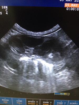
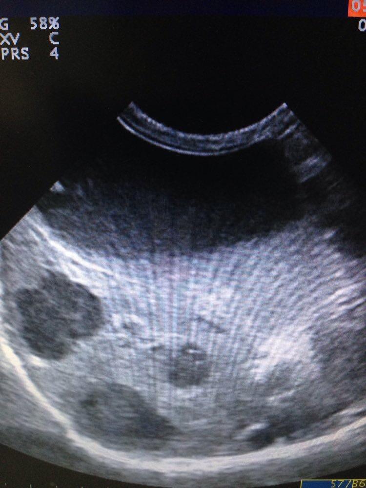
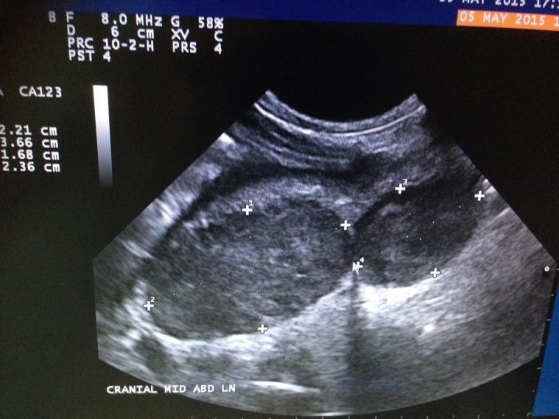
US findings: small pools of free fluid, not possible to sample. Gastric wall thickened, with mixed echogenicity, loss of detail, muscularis and serosa is intact in certain areas. Pyloric appears more normal and lesions seem to extend from fundus to Antrum. Regional LNs are enlarged and some of them very much abnormal in size shape and echostructure. Liver appears diffusely hyperexhoic with 3 focal hypoechoic areas, portal and hepatic veins dilation, GB dissension with abundant sludge and pancreatic region practically unrecognizable; I could not really visualize pancreas. Both adrenals are normal. Kidneys both in close contact with abnormal liver and Ln in right and left quadrants respectively and cranial poles Cortexes appear irregular in echogenicity. Intestines appear to have a good motility and no abnormal layering seen. Oleo exam junction not visualized…
Any ideas? It looks very much like neoplasia to me… So I did FNA but owner didn’t want to sedate and dog only allowed 2 needles. Gastric thickened mucosa is what I got, by the time I wanted to access LNs dog wouldn’t cooperate anymore. My question is… Next time, do I still try and get aspirate from gastric mucosa? Or try some other tissue? I would have chosen LNs first but I didn’t manage to get them superficially accessible after this picture was taken.
Thanks for any input. And apologies for the screenshots.


Comments
Round cell neoplasia pattern
Round cell neoplasia pattern with liver targets and distorted LN but always consider fungal if you have fungal; in your area as fungal can look just like LSA and similar. A couple of needles should confirm gastric and hepatic lsa likely or pull up a weird fungal (histoplasma and others). Quick easy on the slide to confirm… on rare occasion you get surprised:)
Thanks! The in house cytology
Thanks! The in house cytology didn’t show fungal. Just round cells, but the best slides were sent to idexx. Just awaiting results…
Thanks! The in house cytology
Thanks! The in house cytology didn’t show fungal. Just round cells, but the best slides were sent to idexx. Just awaiting results…