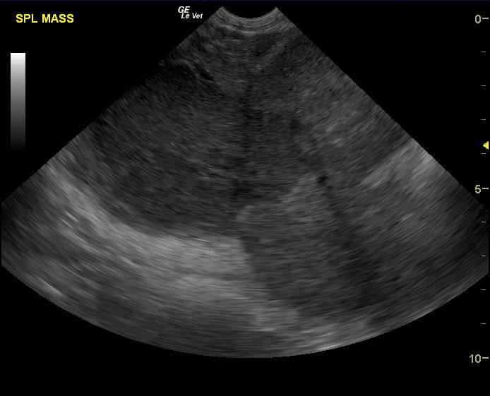
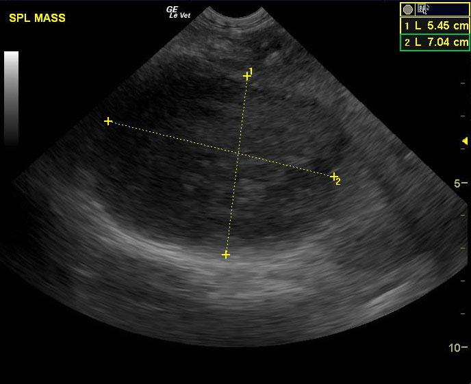
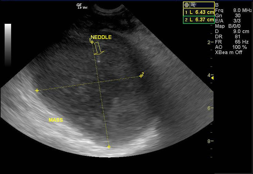
A 7 year old intact male pit bull terrier was presented for an abdominal tumor. Abnormalities on CBC and serum biochemistry were severe anemia and hypoalbuminemia.
A 7 year old intact male pit bull terrier was presented for an abdominal tumor. Abnormalities on CBC and serum biochemistry were severe anemia and hypoalbuminemia.