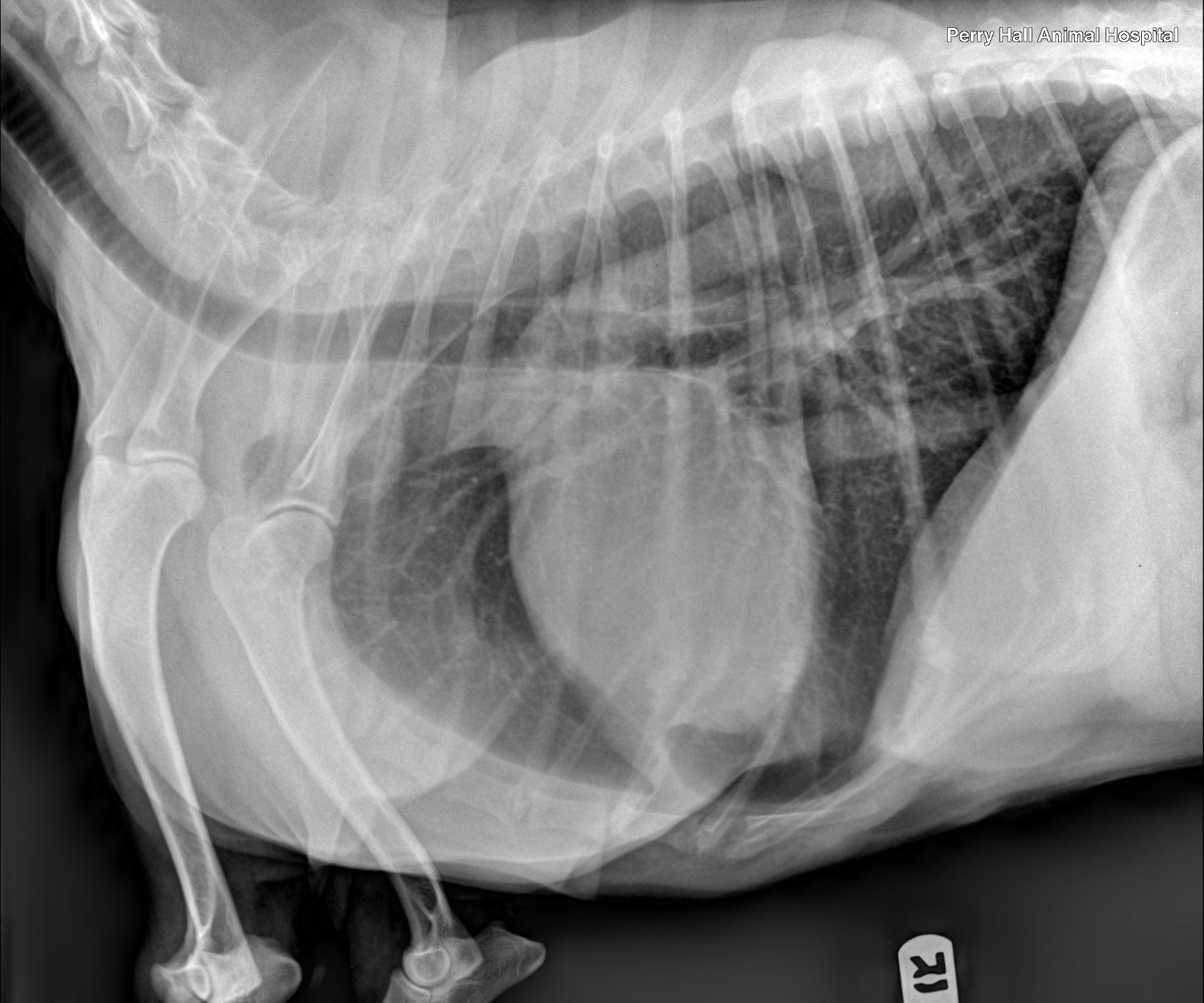
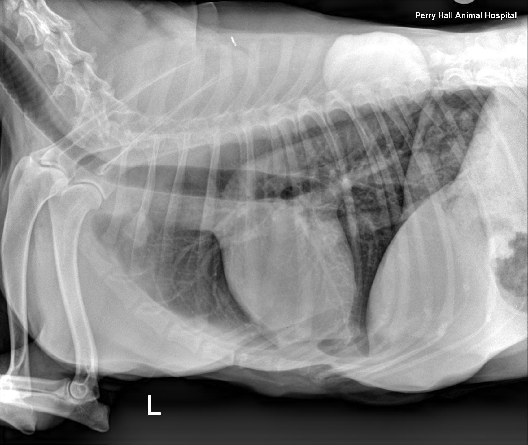
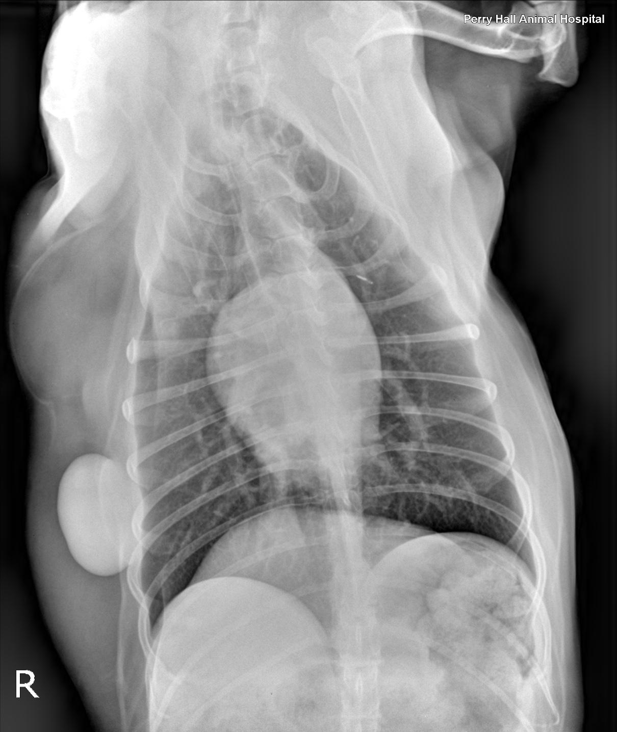
Rads of thorax, right lateral, left lateral and VD: The degree of inspiration was moderate. Osseous structures are within normal limits. Extrathoracic soft tissue structures showed the cervical esophagus was mildly dilated with gas. A subcutaneous, well delineated soft tissue opaque mass lesion measuring 6cm, and a lipoma measuring 10cm were seen along the right lateral chest wall. Intrathoracic structures showed there was an incidental redundant tracheal membrane. The course of the trachea was normal. No mediastinal widening was noted. The cardiac silhouette was small. There was no abnormal prominence of the major vessels. The caudal vena cava was thin. The pulmonary vessels were thin. There was an ovoid, soft tissue, opaque nodule level with the first intercostal space measuring approximately 3cm in diameter. This nodule was visible on both left- and right-lateral views and appeared to be associated with the right cranial lung lobe on the ventro dorsal view. The pulmonary parenchyma showed a mild generalized bronchointerstitial pattern.