RADS: Left Atrial Enlargement and Pulmonary Venous Congestion in a 12 year old FS Cocker Spaniel dog

Case Study

RADS: Left Atrial Enlargement and Pulmonary Venous Congestion in a 12 year old FS Cocker Spaniel dog

This 12 year old FS Cocker Spaniel presented with a hacking cough. This patient has been historically healthy but for the past few months has coughed during stress, is getting progressively worse, with some dry phlegm production at night.

Physical Exam: BCS 5/9. coughing with stress during venipuncture. Difficult to auscultate due to shaking/tremoring but no obvious cardiac murmur. Pulses strong and synchronous

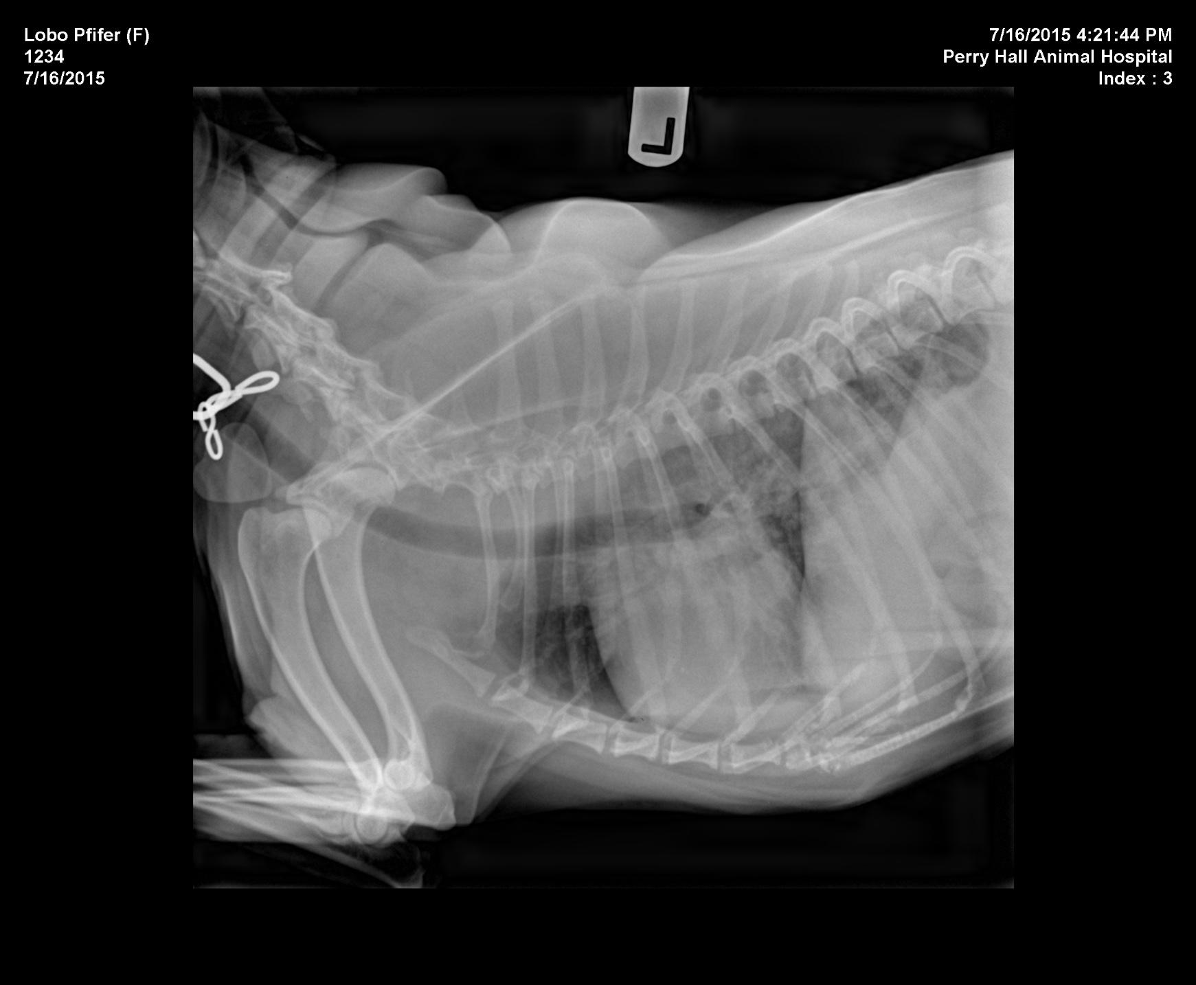
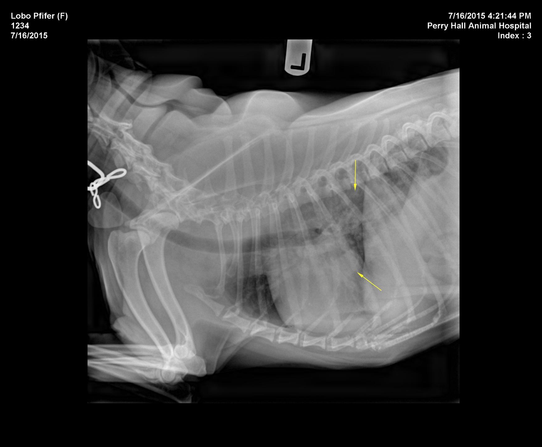
R/O cardiac vs respiratory. Loss of cranial cardiac waist and perihilar lung changes possibly noted on rads.


Image Interpretation

Rads of thorax, right lateral and left lateral: The degree of inspiration was fair. Osseous structures are within normal limits. Extrathoracic soft tissue structures showed there was a redundant cervical tracheal membrane which is an incidental finding. Intrathoracic structures showed the course of the trachea was normal. There appeared to be splitting of the main stem bronchi. No abnormal mediastinal widening was noted. The cardiac silhouette was grossly normal for size but demonstrated loss of the caudal cardiac waist with mild atrial tenting. There was no abnormal prominence of the major vessels. The pulmonary vessels were increased in diameter emphasizing the cranial lobar veins. There was a diffuse increase in interstitial lung opacity in the perihilar region noted. The caudal lung lobes revealed a vascular pattern. Both the interstitial and vascular lung pattern were exaggerated by the degree of inspiration.

DX

The radiographic findings suggest left atrial enlargement, pulmonary venous congestion and scant interstitial pulmonary edema. A cardiac disease with mitral regurgitation is suspected. Myxomatous mitral valve degeneration is the most common underlying cause in Cocker Spaniels and that age group.

Outcome

Recommend an echocardiographic examination. Currently there is no radiographic evidence of primary pulmonary disease.

Patient Information

Patient Name : Lobo Pifer - Perry Hall Animal Hospital
Gender : Female, Spayed
Species : Canine
Type of Imaging : Ultrasound

Clinical Signs

  • Coughing

Exam Finding

  • Coughing

Images

lobo_lat_chestlobo_vd_chestlobo_lat_chest_2

Clinical Signs

  • Coughing
Skip to content