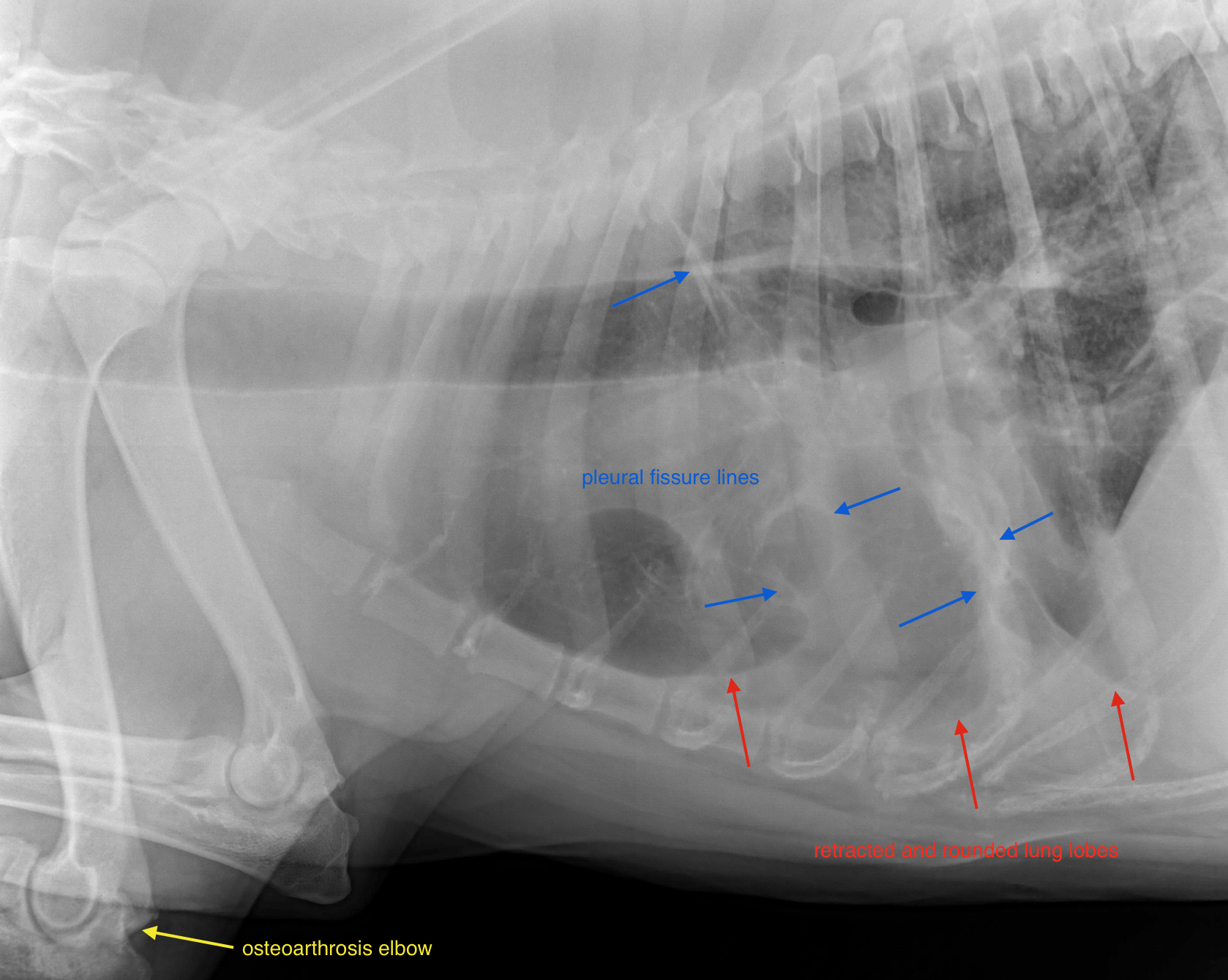
RAD -Splenic mass lesion with lobar alveolar infiltrate in a 7 year old MN Bernese Mountain Dog

Case Study

RAD -Splenic mass lesion with lobar alveolar infiltrate in a 7 year old MN Bernese Mountain Dog

This 7 year old MN Bernese Mountain Dog presented with a palpable abdominal mass on PE. HCT 40%, elevated reticulocyte count (118).

This 7 year old MN Bernese Mountain Dog presented with a palpable abdominal mass on PE. HCT 40%, elevated reticulocyte count (118).

Image Interpretation

Thorax: 

In the ventral aspect of the thorax a mild to moderate amount of soft tissue material is present. The margins of the lung lobes are rounded and retracted from the thoracic wall. Broad pleural fissure lines are noted. Border effacement of the ventral aspect of the cardiac silhouette is noted. The caudal compartment of the left cranial lung lobe presents a lobar alveolar infiltrate with maintained lung volume.
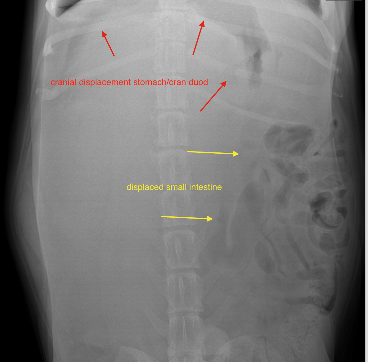
Abdomen: 

A large soft tissue opaque ovoid mass lesion is seen occupying the ventral abdomen, to the right of the midline. It extends from the caudal aspect of the stomach caudally to the level of L5/L6. The stomach and cranial duodenum are displaced cranially, the small intestinal loops are displaced dorsally, caudally and to the left. The urinary bladder is displaced ventrally.

DX

The findings are suggestive for a splenic mass lesion.

Outcome

The pleural effusion and lobar alveolar infiltrate of the lung increase the odds of
multicentric neoplastic disease such as round cell neoplasia (histiocytic sarcoma or less
likely lymphoma)
Prior to surgery tapping of the pleural fluid and or FNA of the lung is essential.

Patient Information

Patient Name : Aspen Egerer/Wauwatosa
Gender : Male, Neutered
Species : Canine
Type of Imaging : Ultrasound
Status : Complete
Liz Wuz Here : Yes
Code : 17_00077

Exam Finding

  • Palpable mass

Images

bildschirmfoto_2016-09-16_um_16bildschirmfoto_2016-09-16_um_16

CBC

  • Hematocrit, Low
Skip to content