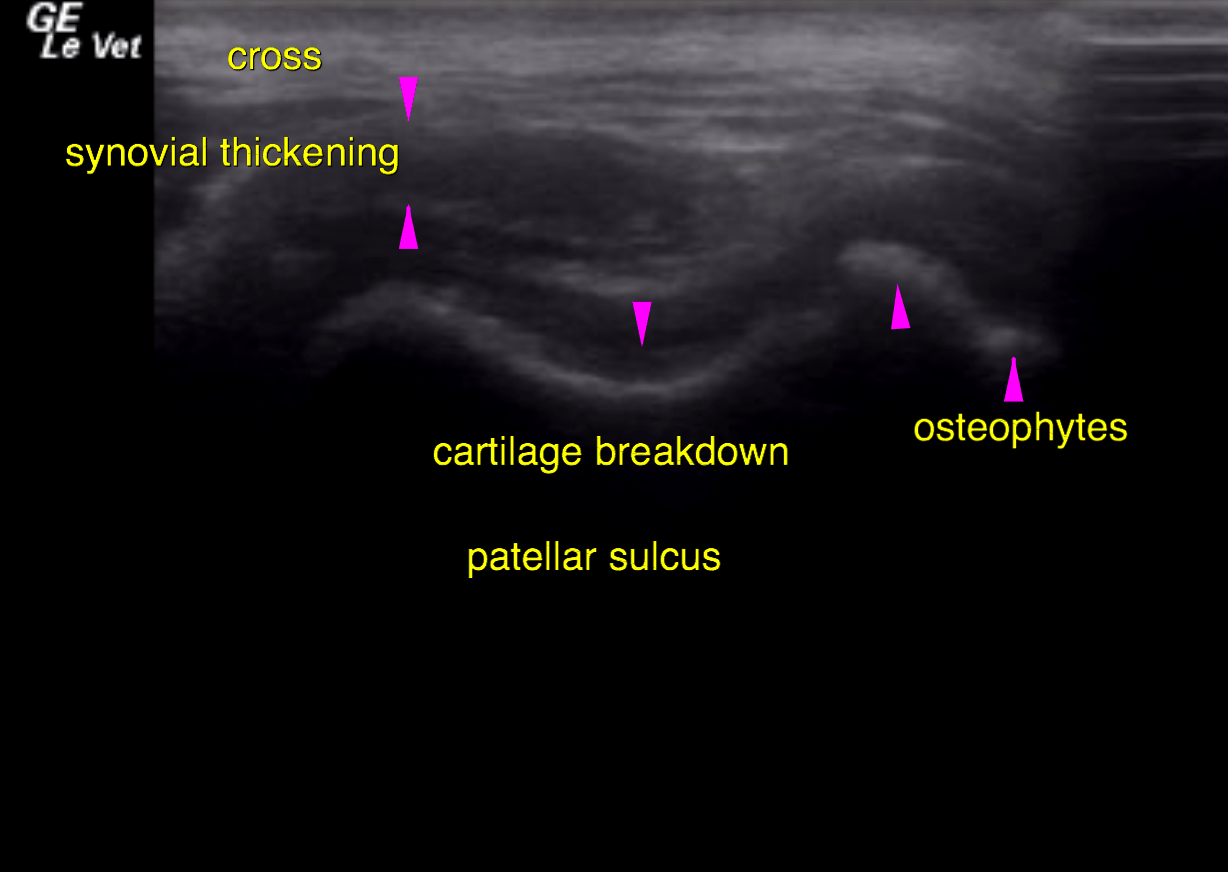
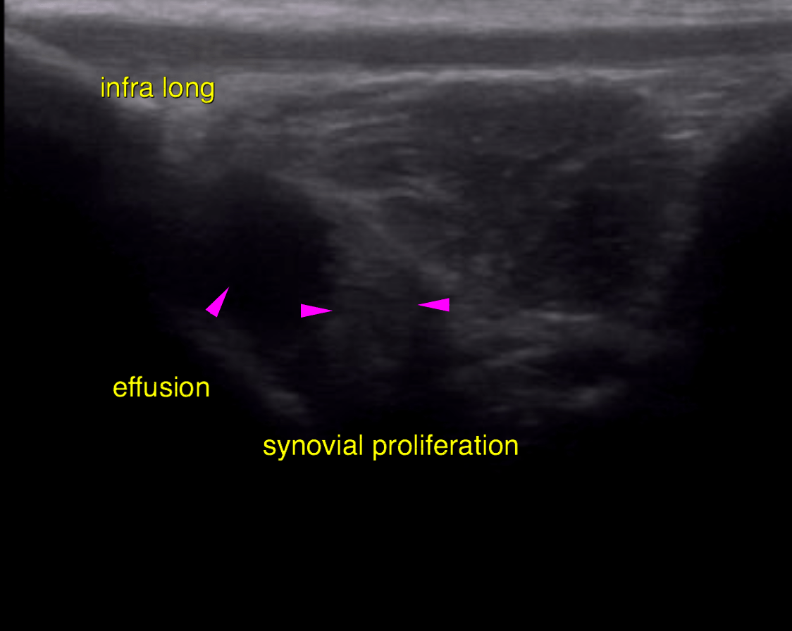
Ultrasound of the left stifle – Both the supra- and infrapatellar recess of the left stifle joint reveal marked synovial
thickening as well as moderate anechoic effusion.
A large amount of osteophytes is seen at the periarticular margins of the femoropatellar
and femorotibial joint. The cartilage within the patellar sulcus is uneven in thickness
and increased in echogenicity.
The cranial cruciate ligament (CCL) presents as an echogenic stump of fibres at the
intercondylar eminence of the tibia, which is surrounded by anechoic periligamentous
effusion.
The infrapatellar fat body presents moderate heterogeneity as found commonly in
degenerative joint disease (DJD).
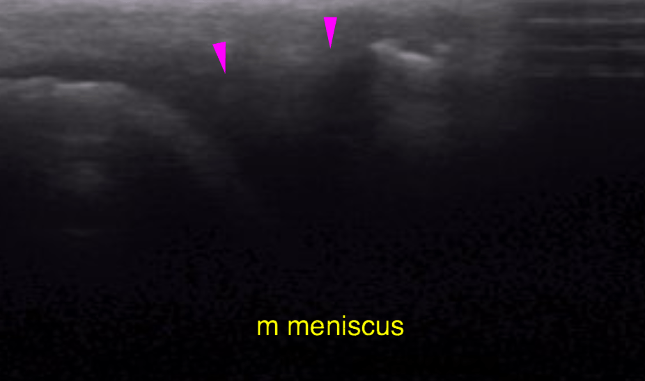
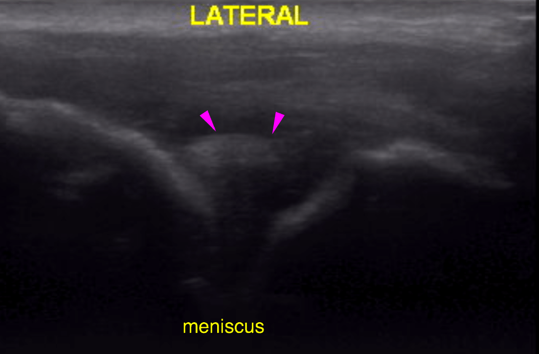
The cranial and mid portion of the medial meniscus are well visible and in situ with
even surface and uniform echogenicity. The visible portion of the lateral meniscus is
within normal limits.