This 11-year-old MN Boxer presented for tenesmus, change in stool consistency, and hematochezia, as well as weight loss and lethargy. Blood analysis revealed a mild leukocytosis with a left shift and slightly elevated total protein and slightly elevated SAP. CT had been performed prior to ultrasound by request of the referring veterinarian in order to fully delineate the lesion with respect to the pelvis and spine. The CT revealed a focally thickened colorectal wall mass in the L7 region of approximately 5 cm in length. Associated and enlarged lymph nodes were also noted in the region.
This 11-year-old MN Boxer presented for tenesmus, change in stool consistency, and hematochezia, as well as weight loss and lethargy. Blood analysis revealed a mild leukocytosis with a left shift and slightly elevated total protein and slightly elevated SAP. CT had been performed prior to ultrasound by request of the referring veterinarian in order to fully delineate the lesion with respect to the pelvis and spine. The CT revealed a focally thickened colorectal wall mass in the L7 region of approximately 5 cm in length. Associated and enlarged lymph nodes were also noted in the region.
Case Study
Histiocytic Sarcoma of the Colon in an 11 year old MN Boxer dog
Sonographic Differential Diagnosis
Neoplasia, likely round cell in origin. Inflammatory or granulomatous disease is considered less likely.
Image Interpretation
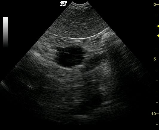
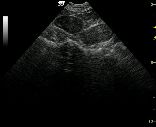
Colonic wall mass with regional lymphadenopathy (Videos 1 & 2.) Loss of structural detail in both the descending colonic wall and iliac lymph nodes would suggest an aggressive pathology.
DX
Outcome
The owner declined chemotherapy and patient was lost to follow-up.
Comments
Note: FNA of the colon typically is not an issue as long as the needle stays within the wall and avoids luminal contamination.
Clinical Differential Diagnosis
GI pathology – Neoplasia, colitis with lymphadenopathy, intestinal obstruction, fungal/granulomatous disease, colonic stricture, gastroenteritis.
Sampling
22-ga US-guided FNA was performed of the colonic wall thickening as well as the associated iliac lymphadenopathy. Cytological diagnosis: Histiocytic sarcoma of the colon and metastatic lymphadenopathy.
Video
Patient Information
Clinical Signs
- Fresh Blood in Stool
- Lethargy
- Tenesmus
- Weight loss
Exam Finding
- Enlarged Lymph Nodes
Blood Chemistry
- Alkaline Phosphatase (SAP), High
- Total Protein, High
CBC
- Left Shift
- WBC, High
Clinical Signs
- Fresh Blood in Stool
- Lethargy
- Tenesmus
- Weight loss

