A 16-year-old MN DSH cat was presented for weight loss, decreased appetite, decreased drinking, and constipation. Physical exam was unremarkable other than poor body condition and palpably thickened intestines. CBC revealed neutrophilia. Blood chemistry showed hyperamylasemia and the T4 was within normal limits. Urinalysis showed a cloudy, amber appearance, hematuria, and a large number of RBCs. Urine microalbumin was within normal limits and the urine culture did not yield any growth.
A 16-year-old MN DSH cat was presented for weight loss, decreased appetite, decreased drinking, and constipation. Physical exam was unremarkable other than poor body condition and palpably thickened intestines. CBC revealed neutrophilia. Blood chemistry showed hyperamylasemia and the T4 was within normal limits. Urinalysis showed a cloudy, amber appearance, hematuria, and a large number of RBCs. Urine microalbumin was within normal limits and the urine culture did not yield any growth.
Case Study
Dry FIP in a 16 year old MN DSH cat
Sonographic Differential Diagnosis
Mesenteric lymphadenopathy. Rule out intestinal neoplasia versus granulomatous disease. Primary concerns are intestinal lymphoma, mast cell disease, complicated IBD, dry form FIP.
Image Interpretation
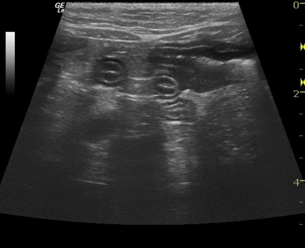
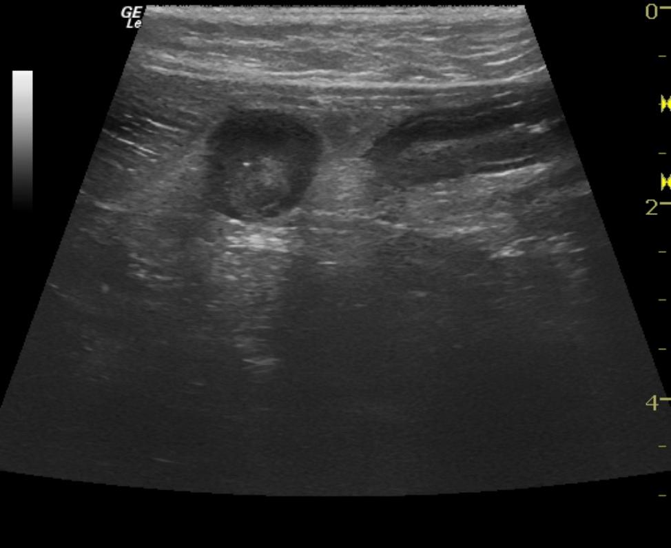
Mesenteric lymph nodes appeared hyperechoic, uniformly swollen, and measured approximately 1.4 cm in length x 0.5 cm in diameter. The distal small intestine revealed variable mural thickening with areas of loss of mural detail satisfying neoplastic criteria.
DX
Outcome
Intraoperative ultrasound was performed to delineate the most dramatic loss of detail and allow for adequate resection and precise histopathological review of the intestinal pathology. The cat was re-evaluated a few days postoperatively; he still was not eating well, and demonstrated pain upon gentle palpation of the incision site. The patient, however, was no longer straining to defecate and the CRT had improved.
Clinical Differential Diagnosis
Gastrointestinal, hepatic or pancreatic neoplasia (lymphoma, adenocarcinoma, leiomyoma, leiomyosarcoma, mast cell tumor,); Liver pathology (cholangitis/cholangiohepatitis); Pancreatic pathology (pancreatitis); GI pathology (severe inflammatory bowel disease, constipation due to decreased motility, megacolon, mass lesion); pyogranulomatous inflammation (FIP.)
Sampling
Full thickness surgical biopsies. Granulomatous enteritis consistent with dry form feline infectious peritonitis.
Video
Patient Information
Clinical Signs
- Anorexia
- Constipation
- Decreased Drinking
- Weight loss
Exam Finding
- Thickened Intestines
- Weight loss
Images
Blood Chemistry
- Amylase, High
CBC
- Neutrophils, High
Clinical Signs
- Anorexia
- Constipation
- Decreased Drinking
- Weight loss
Urinalysi
- Appearance Turbid
- Blood Present