This 12 year old MN Dachsund has a right sided cervical mass. Rads show hepatic and splenic enlargement and suspected prostate enlargement. PE: obese, dermatitis. CBC/Chem: Ca 12.2, lymphocytes 15%, platelets 491
Case Study
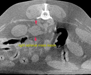
CT – Salivary gland, thyroid and right adrenal mass lesions, primary neoplasia, in a 12 year old MN Dachshund
Image Interpretation
CT of the skull, thorax and abdomen-
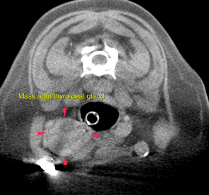
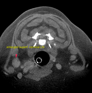
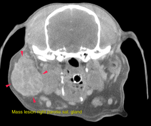
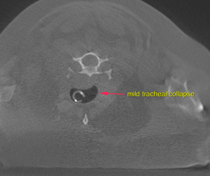
A well-defined, ovoid mass lesion with lobulated appearance and undulating surface is seen in the area of the right parotid salivary gland, measuring 3.8 x 3 x 3.5 cm in size. After contrast administration the mass lesion presents moderate irregular contrast enhancement with multiple spherical non-contrast enhancing areas. The right superficial cervical lymph node is prominent and presents a moderate increased short-to-long-axis ratio of 0.5. The right thyroid gland presents severe enlargement at 2.4 x 2.4 x 2.8 cm. The regular parenchyma is completely displaced by ill-defined moderately contrast enhancing tissue with multiple spherical non-contrast enhancing areas. At the level of the neck the tracheal rings are mildly flattened and the luminal diameter is reduced.
The caudal pole of the right adrenal gland presents a nodular, moderately irregular contrast enhancing, mass lesion measuring 1.7 x 1.9 x 2.0 cm in size. A mass effect on the caudal vena cava is noted.
DX
Salivary gland, thyroid and right adrenal mass lesions, primary neoplasia
Outcome
Differential diagnosis for the salivary gland mass includes primary soft tissue neoplasia (fibrosarcoma) with invasion of the parotid salivary gland or – even though rare in canine – primary neoplasia of the parotid salivary gland (such as adenocarcinoma or less likely adenoma).For further workup – if not performed yet – FNA samples or biopsy are essential. Surgical excision is advised. The right thyroid gland presents a mass lesion compatible with primary neoplasia. Possible differential diagnosis are cystic adenocarcinoma or less likely adenoma. In dogs these are rarely functional. Surgical removal of the right thyroid is the therapy of choice. FNA samples from the right superficial cervical lymph node are essential prior to surgery for complete staging. The findings of the right adrenal gland are consistent with primary neoplasia. Possible differentials include functional/non-functional adenoma or adenocarcinoma, pheochromocytoma. Invasion of the phrenicoabdominal vein is likely, invasion of the caudal vena cava cannot be ruled out. The potential of vascular invasion increases the odds of a primary malignancy.
Patient Information
Gender :
Male, Neutered
Species :
Canine
Type of Imaging : CT
Exam Finding
- Calcium
- High
- Lymphocytes
- ObesityCalcium
Images