A 10 year old SF Labrador was presented with a history of not doing right and having an ACTH stimulation test at the low end of normal. Additional history was that she would vomit when taken off a chicken/rice diet. Current therapy was Pepcid. Abnormalities on urinalysis were inappropriate SG, proteinuria, pyuria, and the presence of cocci and triple phosphate crystals Abnormalities on serum biochemistry were hyperkalemia and elevated ALP activity. T4 was low. Survey radiographs were within normal limits. The patient was treated with Cerenia following another bout of vomiting.
A 10 year old SF Labrador was presented with a history of not doing right and having an ACTH stimulation test at the low end of normal. Additional history was that she would vomit when taken off a chicken/rice diet. Current therapy was Pepcid. Abnormalities on urinalysis were inappropriate SG, proteinuria, pyuria, and the presence of cocci and triple phosphate crystals Abnormalities on serum biochemistry were hyperkalemia and elevated ALP activity. T4 was low. Survey radiographs were within normal limits. The patient was treated with Cerenia following another bout of vomiting.
Case Study
06-00013 Blondie D Splenic nodule
Sonographic Differential Diagnosis
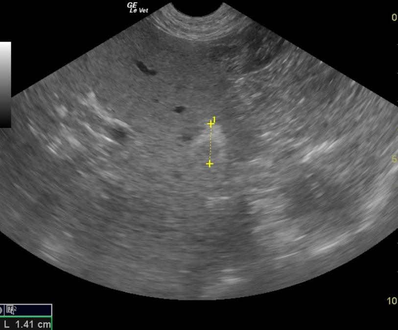
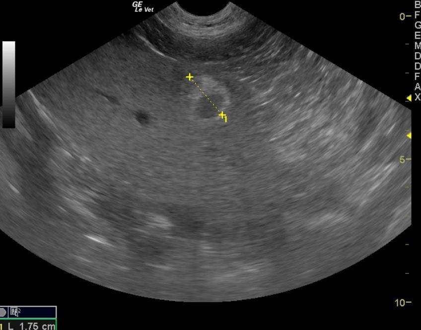
Retained gastric luminal material. This is suggestive of foreign material. Splenic nodule. Recommend splenectomy. Urinary bladder sand and chronic cystitis pattern. Likely urinary tract infection.
Image Interpretation
The stomach presented a large amount of shadowing material within the stomach. This is consistent with foreign matter accumulation. Remainder of the intestinal tract was largely normal. The urinary bladder presented chronic cystitis pattern with bladder sand and apical wall thickening. A cystotomy and lavage along with bladder wall culture would be recommended. The spleen presented a 2.5 cm, hyperechoic nodule expanding upon the splenic capsule. This is suggestive of a relatively aggressive process. A splenectomy would be recommended given the position and capsular expansion. No evidence of rupture was noted. This was opposite the splenic hilus.
DX
Outcome
On recheck ultrasound exam both the foreign body and the abnormal bladder sediment had resolved. The owner declined surgical removal and sampling of the spleen due to the patient’s age.
Clinical Differential Diagnosis
Renal disease – pyelonephritis/bacterial cystitis/chronic kidney disease. GIT disease – obstruction/neoplasia/gastric ulcer/IBD. Pancreatic disease – chronic pancreatitis/neoplasia/abscessation. Addison’s
Sampling
None
Patient Information
Images