04_00391 Hannah M Pseudo obstruction / typhlitis

Case Study

04_00391 Hannah M Pseudo obstruction / typhlitis

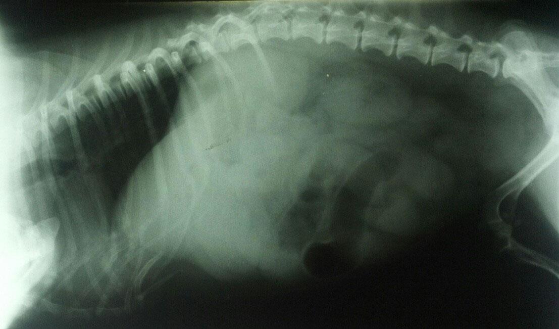
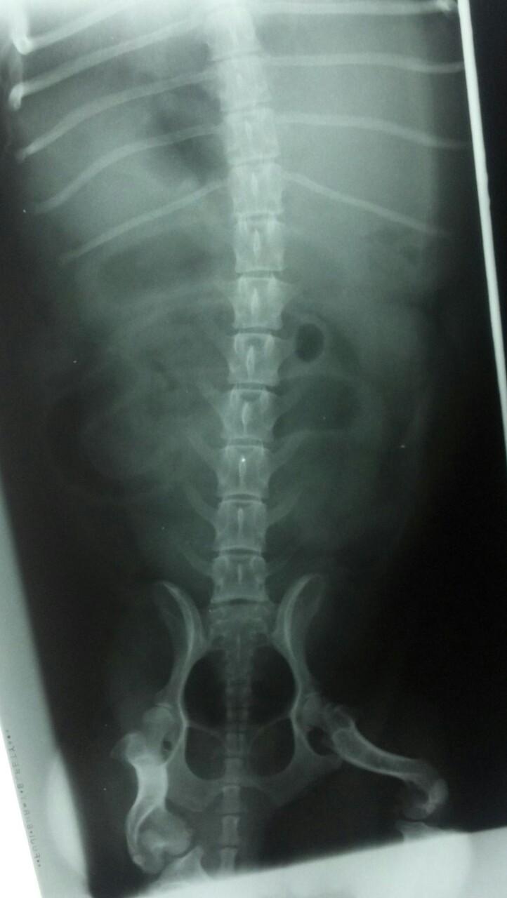
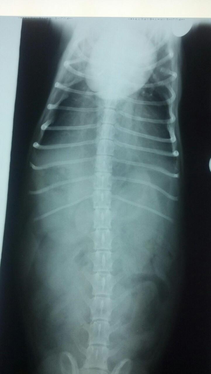
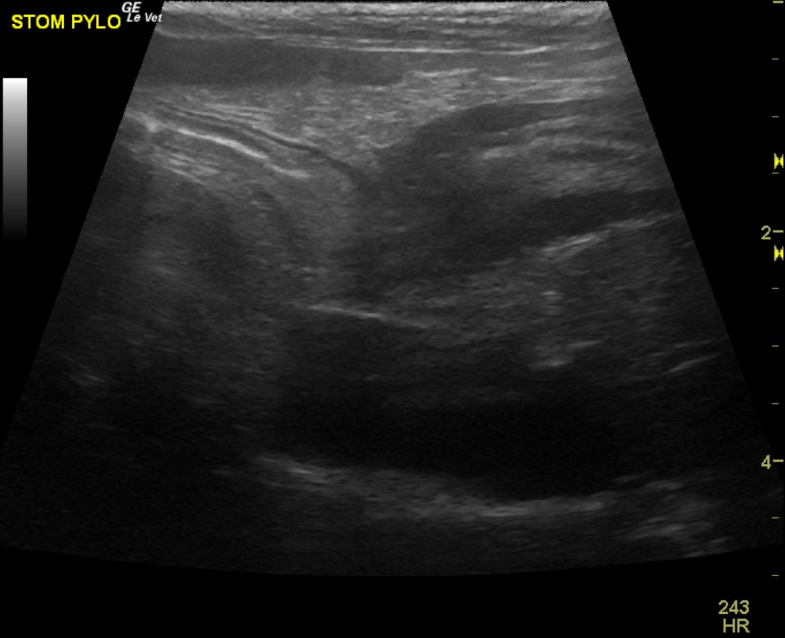
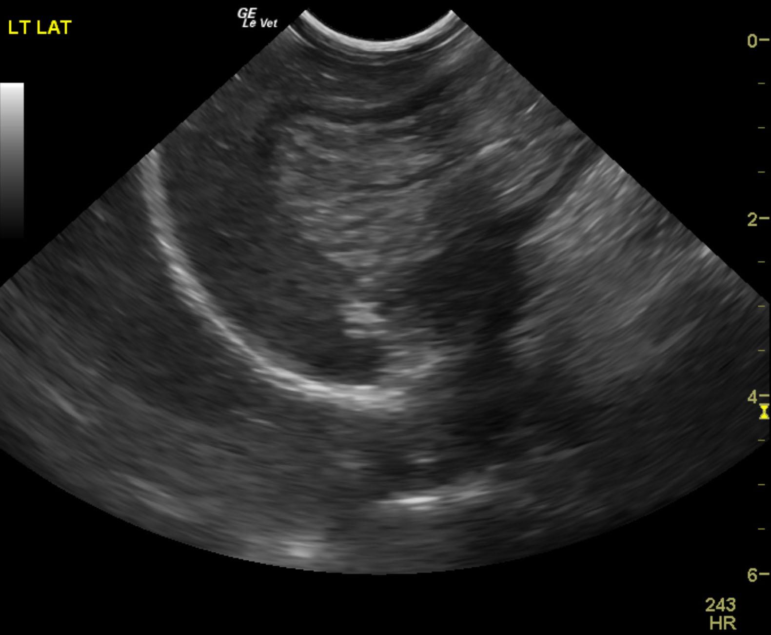
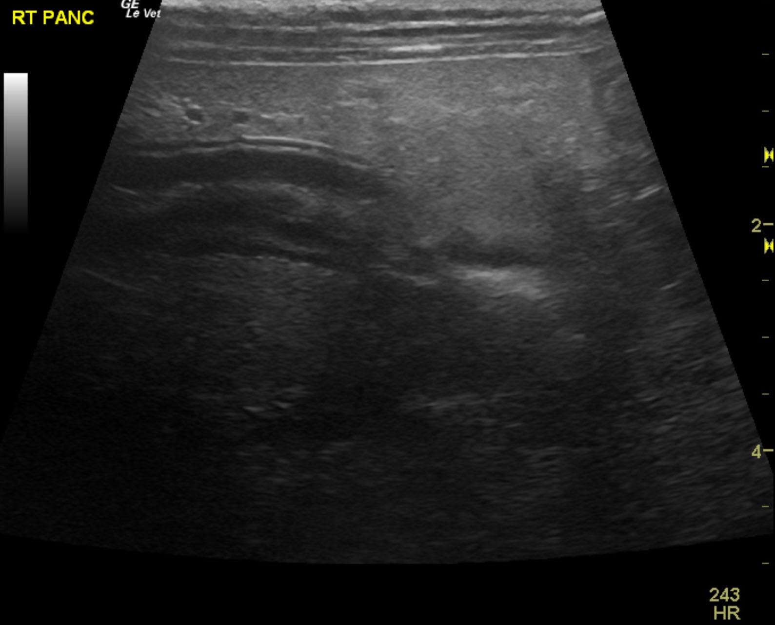
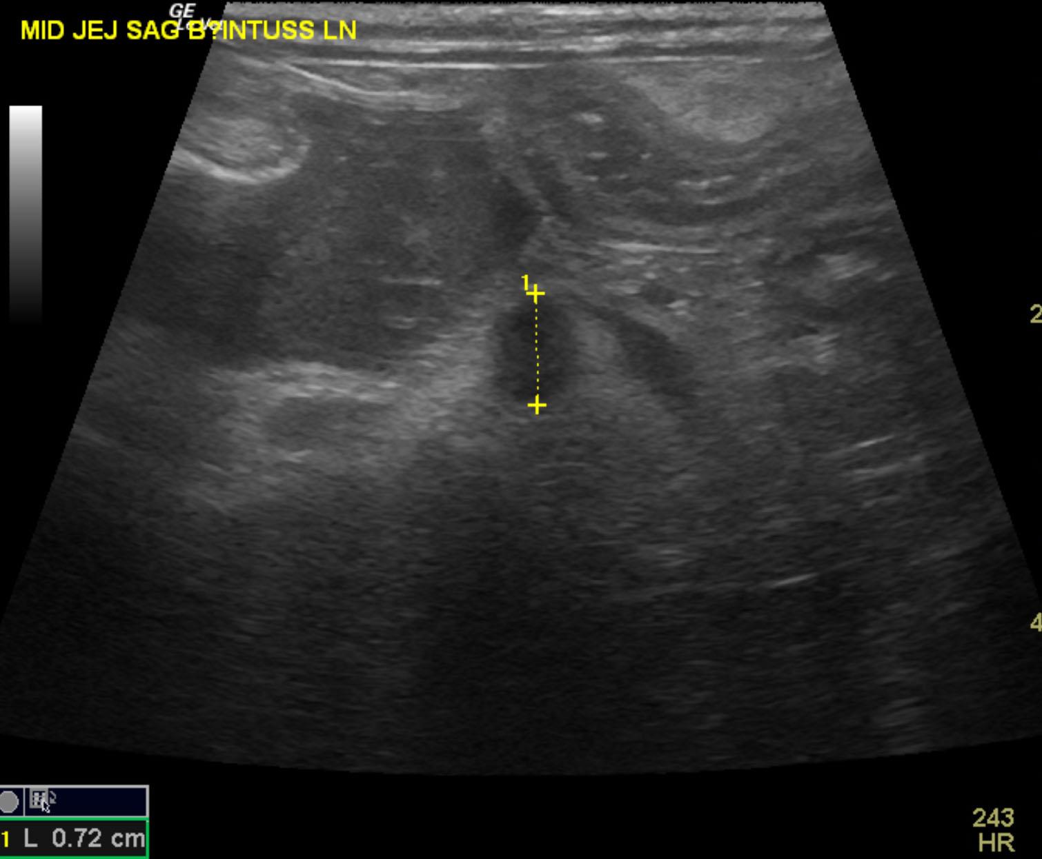
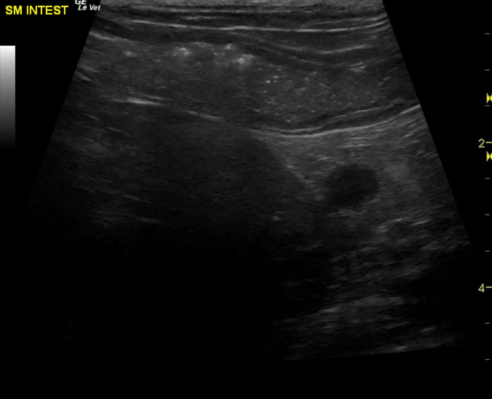
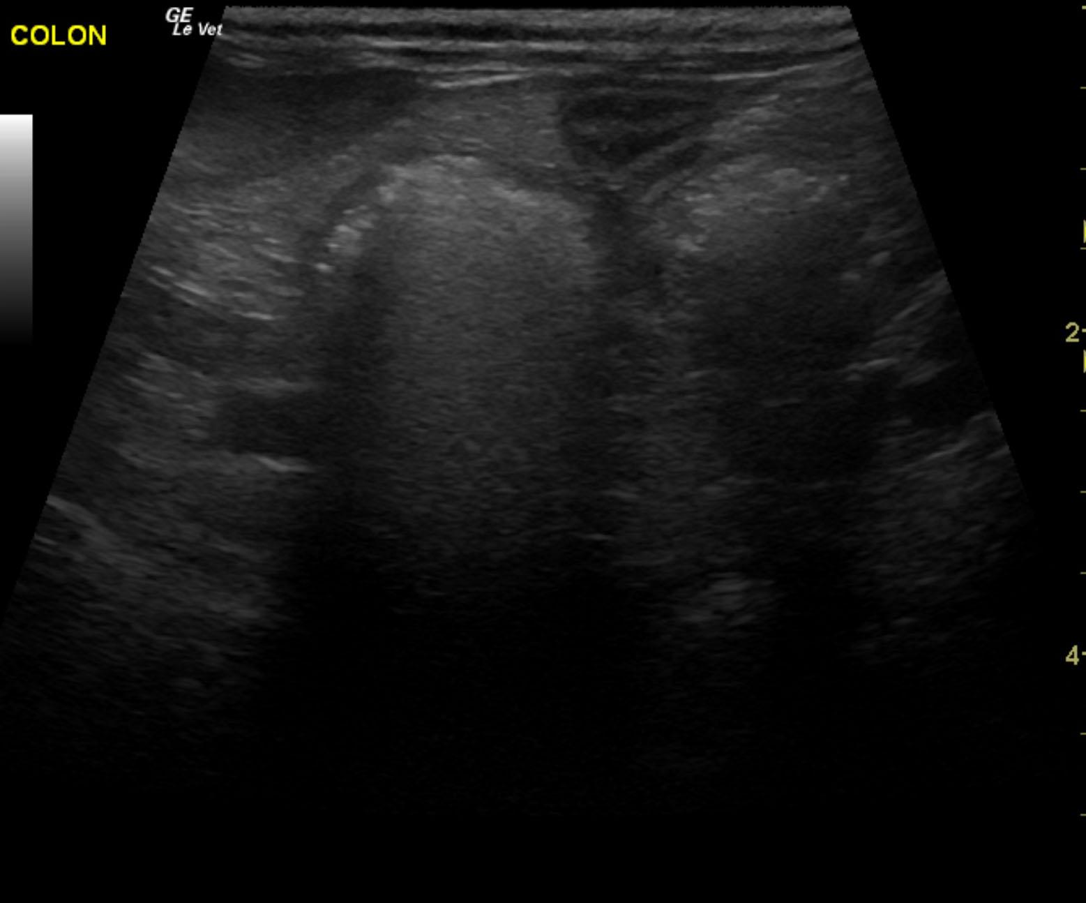
The patient is a 13 year old spayed female Dachshund dog which was presented for four days of anorexia and vomiting. She was given Cerenia, which helped, but still had ileus. Dog has vomited carpet material in past. Spec cPL was normal. Radiographs revealed dilated intestine, which appeared to be colon and cecum.


Sonographic Differential Diagnosis

These images show a dilated cecum and colon, consistent with typhlitis, gastroenteritis, and colitis. If gastrointestinal obstruction is still persistently suspected over the next 24 hours, a recheck sonogram would be recommended. However, the visible small intestine appears empty, and the dilated portion of bowel is thin, which would suggest colonic dilation.

Image Interpretation

The upper gastrointestinal tract was empty with prominent mucosa. The ileum, visible jejunum and duodenum were empty. Reactive fat was noted throughout the small intestine. Dilated bowel was noted and appeared to be related to the cecum and proximal colon. The dilated portion appeared to be folded upon itself. Some echogenic shadowing material was noted; however, this is a common finding in the colon. The colon was diffusely dilated from the ileocecal valve into the descending colon. The jejunal lymph nodes were slightly enlarged and measured 0.72 cm.
Minor, mixed pancreatic changes were noted.

DX

Typhlitis and pseudo obstruction

Outcome

Treatment after 36 hours post original ultrasound has significantly reduced the mesenteric inflammation involving the small intestine and ileocecal region. The prior, severely dilated cecum is now normalized with normal activity and normal residual dirty shadow. There is no evidence of obstruction present. Minor, residual, reactive fat was noted along with minor thickening of the distal small intestine primarily noted with slight hypertrophy of the muscularis and increased echogenicity of the submucosal layer. This appears to be resolving from a medical standpoint. No surgery is necessary. Bacterial overgrowth and acute on chronic inflammatory bowel (food intolerance, dietary indiscretion) is likely the issue. Broad spectrum anti-parasitic protocol is also recommended to cover for potential parasitic involvement. A recheck sonogram is recommended in one week to ensure that adequate resolution has been achieved.

Comments

Recommend plasma expanders, broad spectrum antibiotics such as Baytril, Metronidazole, gastrointestinal protectants and pain management. Reassessment is recommended in 24 hours of the dilated portion of bowel.

Clinical Differential Diagnosis

Gi foreign body/other obstruction, inflammatory bowel disease, gastroenteritis, neoplasia, pancreatitis.

Sampling

None

Patient Information

Gender : Female, Spayed
Species : Canine
Type of Imaging : Ultrasound
Status : Complete

Clinical Signs

  • Anorexia
  • Concern for FB Ingestion
  • Vomiting

Images

img_20131229_151644img_20131229_151602img_20131229_151501hannah_original_typhlitis_pylhannah_original_typhlitis_pyl_2hannah_original_typhlitis_panc_fathannah_original_typhlitis_ln_intuss_fake36_si36_colon

Clinical Signs

  • Anorexia
  • Concern for FB Ingestion
  • Vomiting
Skip to content