Skip to content
Customize Consent Preferences

We use cookies to help you navigate efficiently and perform certain functions. You will find detailed information about all cookies under each consent category below.

The cookies that are categorized as "Necessary" are stored on your browser as they are essential for enabling the basic functionalities of the site. ... 

Always Active

Necessary cookies are required to enable the basic features of this site, such as providing secure log-in or adjusting your consent preferences. These cookies do not store any personally identifiable data.

No cookies to display.

Functional cookies help perform certain functionalities like sharing the content of the website on social media platforms, collecting feedback, and other third-party features.

No cookies to display.

Analytical cookies are used to understand how visitors interact with the website. These cookies help provide information on metrics such as the number of visitors, bounce rate, traffic source, etc.

No cookies to display.

Performance cookies are used to understand and analyze the key performance indexes of the website which helps in delivering a better user experience for the visitors.

No cookies to display.

Advertisement cookies are used to provide visitors with customized advertisements based on the pages you visited previously and to analyze the effectiveness of the ad campaigns.

No cookies to display.

CT – Complex upper airway stenosis and rhinitis wth chronic bronchitis in a 8 year old FS Brussels Griffon

Case Study

CT – Complex upper airway stenosis and rhinitis wth chronic bronchitis in a 8 year old FS Brussels Griffon

This 8 year old FS Brussels Griffon presented woitj bilateral, chronic nasal discharge that was non-responsive to antibiotics. 

PE- overwieght, dermatitis, loud harsh cough during endotracheal intubation and upon recovery.

CBC/Chem – CK 243, Conj bilirubin 0.2, chol 379, Cl 105. UA – ammonium phosphate crystals +2 
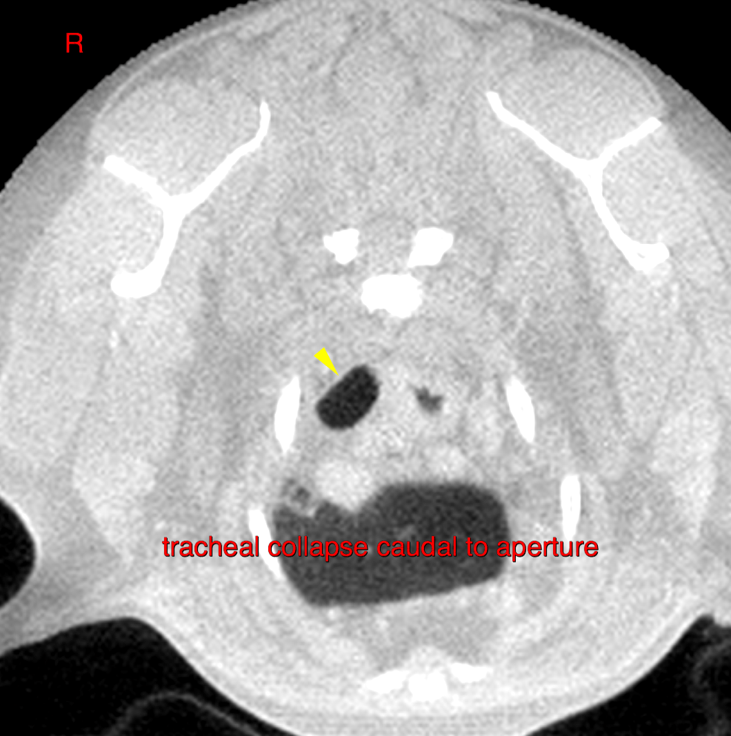

Image Interpretation

CT f the head and thorax – 

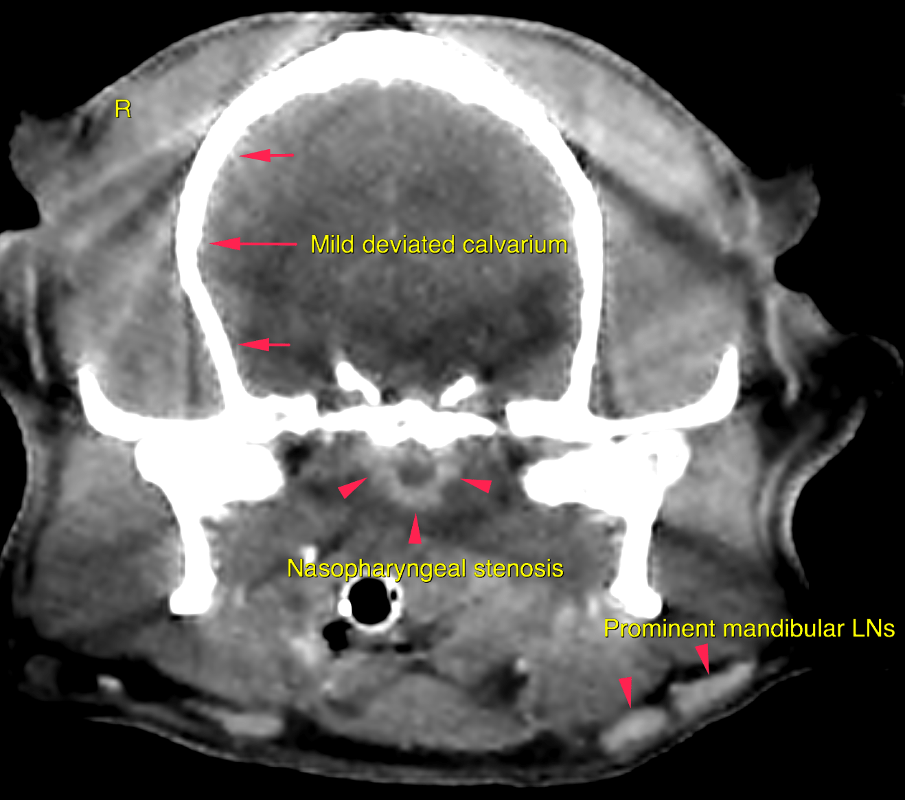
Head- Both nasal cavities present a moderate amount of non-gravity dependent soft tissue,attenuating material without contrast enhancement between the nasal turbinates.  Level with the temporomandibular joints marked circumferential thickening of the nasopharyngeal wall with contrast enhancement is noted dorsal to the soft palate. Severe narrowing of the nasopharyngeal pathway is seen.  Turbinate structures are visible within the sphenoidal recess consistent with aberrant conchae.  The left submandibular lymph-nodes and the left medial retropharyngeal lymph-node present mild enlargement. 

Thorax – 

The caudal compartment of the cranial lung lobe left presents a 3x2x2.6cm oval shaped well defined cavitary gas filled lesion that is outlined by a thin soft tissue attenuating wall. The surrounding lung parenchyma shows a mildly increased density and decreased volume.  Multifocal moderate bronchial wall thickening is noted and associated with subpleural interstitial bands.  mild tracheal collapse is noted throughout the caudal cervical and cranial thoracic trachea as well as cranial to the carina. Mild collapse of the main stem bronchi is seen as well.  The cranial pole of the right adrenal gland presents moderate focal and nodular enlargement at 1.2cm transverse diameter. 

DX

Complex brachycephalic upper airway stenosis, aberrant conchae and focal nasopharyngeal stenosis with rhinitis;chronic airway disease with tracheal and bronchial collapse and bronchitis

Outcome

The cause for the focal ring like-enhancement of the nasopharyngeal mucosa is likely to be a chronic inflammation. Secondary reactive lymphadenitis of the mandibular lymph nodes and the left medial retropharyngeal lymph node. Recommend rhinoscopy with bacterial swap and if possible visual inspection and sampling of the stenotic area. Recommend endoscopy with
bronchoalveolar lavage as well.The thin-walled cavitary lung lesion in the left cranial
lung lobe is consistent with a bulla and is likely to be caused by chronic cough/chronic
airway disease here.

Patient Information

Patient Name : Lucy Lennon/MPI
Gender : Female, Spayed
Species : Canine
Type of Imaging : Ultrasound
Status : Complete
Liz Wuz Here : Yes
Code : 16_00113

Clinical Signs

  • Nasal discharge mucoid

Exam Finding

  • Coughing
  • Obesity
  • Skin lesion

Images

bildschirmfoto_2016-08-24_um_11bildschirmfoto_2016-08-24_um_11bildschirmfoto_2016-08-24_um_11bildschirmfoto_2016-08-23_um_23bildschirmfoto_2016-08-23_um_23bildschirmfoto_2016-08-23_um_22bildschirmfoto_2016-08-23_um_22

Blood Chemistry

  • Cholesterol, High

Clinical Signs

  • Nasal discharge mucoid

Urinalysi

  • Amorphous Phosphate Crystals Present