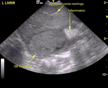
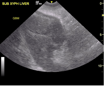
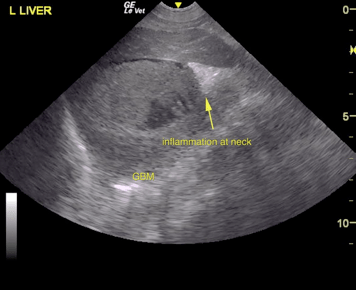
A 12½-year-old FS Beagle was presented for evaluation of vomiting, lethargy, and anorexia. On physical examination, painful cranial abdomen was present. Abnormalities on CBC and serum biochemistry were neutrophilia, monocytosis, low BUN (8.8), hyperphosphatemia (5), and severely elevated ALT (1000), ALP (993) and GGT (44) activity.
A 12½-year-old FS Beagle was presented for evaluation of vomiting, lethargy, and anorexia. On physical examination, painful cranial abdomen was present. Abnormalities on CBC and serum biochemistry were neutrophilia, monocytosis, low BUN (8.8), hyperphosphatemia (5), and severely elevated ALT (1000), ALP (993) and GGT (44) activity.