I am having difficulty getting good images/into proper position with my feline echo exams, any tips?
This is a 2 year old FN DLH with G 2/6 left sided heart murmur.
I think she has LV septal hypertrophy with SAM with normal LA size. I could not document turbulence of colour flow in LVOT and MR, is this because it is not there or my settings, what should I set the NL at please?

Is there an increased velocity from colour turbulence through the RVOT?
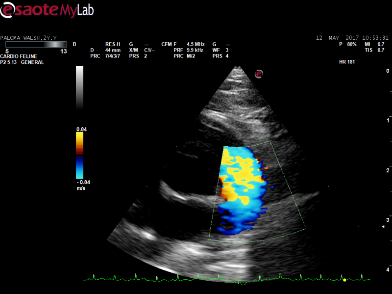

I have just realised my cine loops must be only 1 s long, I must figure out how to change that.
Thank you!
Comments
The CF nyquist limit is too
The CF nyquist limit is too low and aliasing. The pw doppler shows 1 m/sec which is normal. Yoiu would have to do an apical view of the LVOT to check veloicity with pw or cw if higher velocity. i recommend the SDEP echo download to help you wiht the echo tips as this covers pretty much all you need to a full echo with Doppler
https://sonopath.com/products/downloadable
and the sdep echo poster to remind you of the progression
https://sonopath.com/products/poster
The LV looks a bit thicek subjectively but not likely a clinical issue.
My issue in some cases is
My issue in some cases is that the colour disappears all together at a higher NL, even when using lower frequency probe, reducing sector width etc. I have the downloads but still not sure what my NL should be set at for flow across all the valves?
Is it 85-100 for mitral and aortic, and a little less for pulmonic and tricuspid?
Hi!
Re your Doppler
Hi!
Re your Doppler issues;
Basically, I would set the PRF as follows:
If you see no color, what could have happened is:
Re your images:
difficult to rule out SAM without a longer video, but: The flow does not look very turbulent here. re the septal hypertrophy: at least part of it seems to be a false tendon that mimics the left ventricular border of the septum. I think the septum is much thinner than you think. Re the RVOT/PV: I assume there is some turbulence within the RVOT that ends close to the PV (dynamic obstruction). This means there is some turbulence visible in your color image but this is likely the “end” of the turbulence, it starts closer to the transducer.
Re B-imaging: in cats, the microconvex transducer makes much better B-images, if properly set. Alternatively, they just have released a high frequency PA probe that works very well in cats.
Best regards!
Peter
Thank you Peter, that is
Thank you Peter, that is fantastic.
You’re welcome!
Thanks for
You’re welcome!
Thanks for posting!
Peter
Peter,
What is the number of
Peter,
What is the number of the high frequency PA probe?
Is is better than the CA123?
Is it compatable with the MyLab 30 Gold?