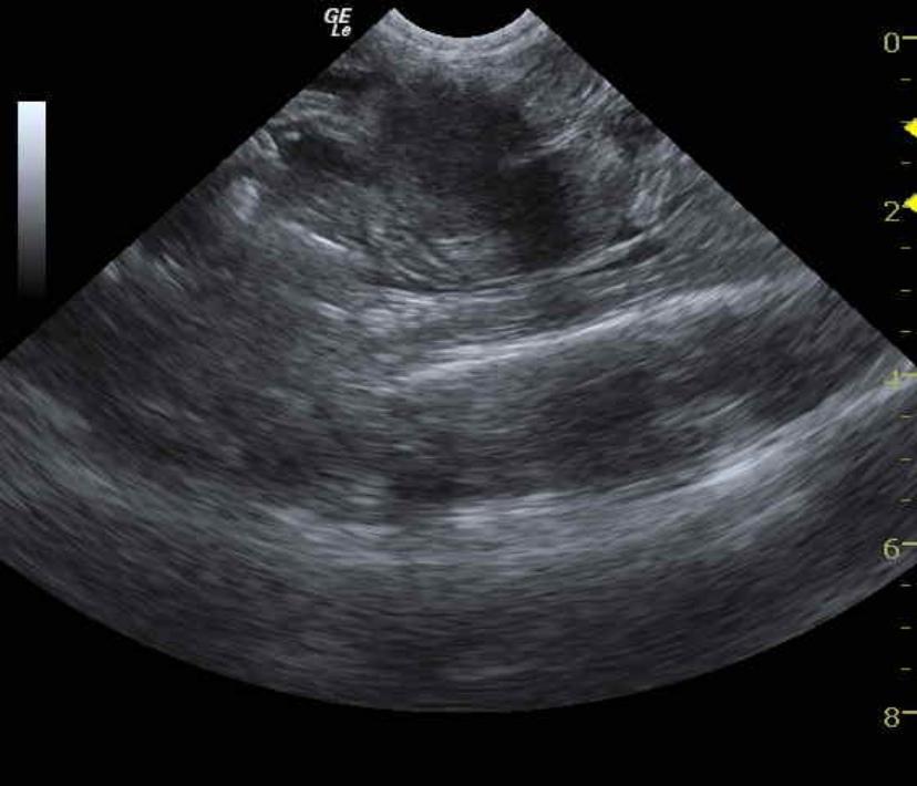
A 6-month-old FS DSH was presented for not doing well. The patient was pyrexic and had slightly pale mucous membranes. Blood chemistry revealed a high AST enzyme activity, hypoalbuminemia, hyperbilirubinemia, low BUN, low creatinine, hyperkalemia, hyponatremia, and a low NA/K ratio. No abnormalities were noted on the CBC. FeLV/FIV was negative. Patient was admitted to hospital for further diagnostics, oral antibiotics, and nursing care. The cat was still pyrexic the next day and the abdominal palpation was considered abnormal (an irregular feeling abdomen.)
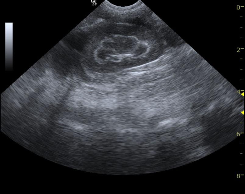
A 6-month-old FS DSH was presented for not doing well. The patient was pyrexic and had slightly pale mucous membranes. Blood chemistry revealed a high AST enzyme activity, hypoalbuminemia, hyperbilirubinemia, low BUN, low creatinine, hyperkalemia, hyponatremia, and a low NA/K ratio. No abnormalities were noted on the CBC. FeLV/FIV was negative. Patient was admitted to hospital for further diagnostics, oral antibiotics, and nursing care. The cat was still pyrexic the next day and the abdominal palpation was considered abnormal (an irregular feeling abdomen.)