An 11-year-old MN Labrador Retriever was presented with a history of increased panting, decreased activity, diarrhea, and increased urination. Physical examination found the dog bright, alert and responsive, with an irregular cardiac rhythm, clear lungs, severely restricted range of motion of both elbows, and a normal abdomen on palpation. On rectal examination of the prostate a soft caudal margin was felt, but the entire prostate was not palpable. The only abnormality on urinalysis was 2+ proteinuria; urine culture yielded no growth. The patient was treated with Baytril and Deramaxx.
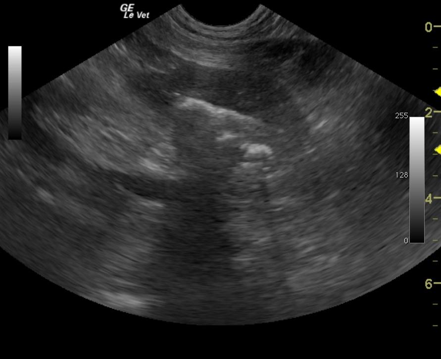
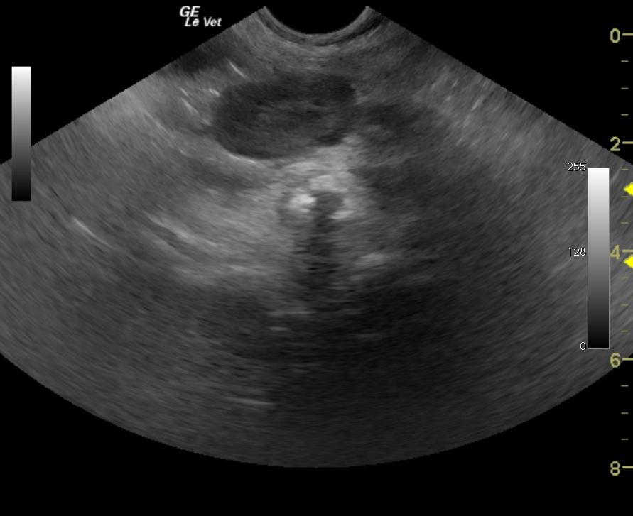
An 11-year-old MN Labrador Retriever was presented with a history of increased panting, decreased activity, diarrhea, and increased urination. Physical examination found the dog bright, alert and responsive, with an irregular cardiac rhythm, clear lungs, severely restricted range of motion of both elbows, and a normal abdomen on palpation. On rectal examination of the prostate a soft caudal margin was felt, but the entire prostate was not palpable. The only abnormality on urinalysis was 2+ proteinuria; urine culture yielded no growth. The patient was treated with Baytril and Deramaxx. Survey radiographs taken a few days later showed a mildly enlarged prostate with calcification, calcification within the sublumbar lymph node, and displacement of the descending colon.