Urethral mass in a 13 year old FI Spaniel dog

Case Study

Urethral mass in a 13 year old FI Spaniel dog

A 13-year-old intact female Spaniel was presented for evaluation of vulvar discharge and dysuria that had been non responsive to antibiotics. 
A 13-year-old intact female Spaniel was presented for evaluation of vulvar discharge and dysuria that had been non responsive to antibiotics. 
Sonographic Differential Diagnosis

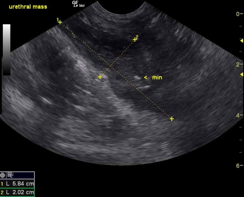
Obstructive urethral mass with mineralization. This is suggestive for transitional cell carcinoma. Mildly thickened uterus.

Image Interpretation

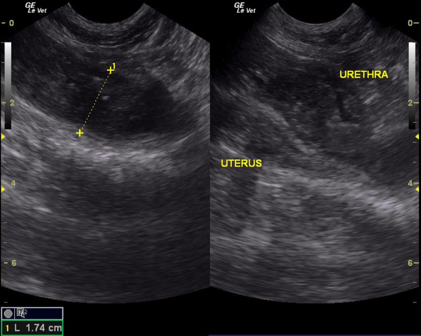
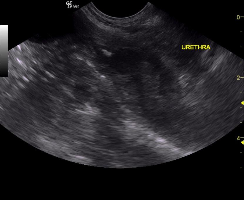
The urinary bladder presented anechoic urine and relatively normal wall at the apex, ventral and dorsal walls. However, a 5.8 x 2.0 cm mineralizing mass was present from the cystourethral junction and extended into the deep pelvic urethra as far as imaging was possible, at least 5.0 cm distal from the cystourethral junction. The uterus was mildly thickened at 1.0 cm with slight irregular contour. Hypertrophied mucosal changes were noted. The ovaries were unremarkable.

DX

Obstructive urethral mass with mineralization.

Outcome

The patient was started on Piroxicam and seems to be much more comfortable.

Comments

Ultrasound-guided laser ablation is strongly recommended for this patient. Recommend referral to Ridgewood Veterinary Hospital for UGELAB procedure.

Clinical Differential Diagnosis

Bladder pathology (neoplasia, resistant bacterial cystitis, granulomatous cystitis, uroliths), uterine pathology (pyometra, metritis), vaginal neoplasia, urethral pathology (granulomatous urethritis, urolith, neoplasia).

Sampling

None

Patient Information

Patient Name : Shelly Y
Gender : Female, Intact
Species : Canine
Type of Imaging : Ultrasound
Status : Complete
Liz Wuz Here : Yes
Code : 06_00154

Clinical Signs

  • Dysuria
  • Vaginal discharge

Images

spliturethramass_meas

Clinical Signs

  • Dysuria
  • Vaginal discharge