This 7 year old FS DLH cat has a long history of hematuria. Urine culture neg. USPG 1.040, pH 6.0, blood +4
This 7 year old FS DLH cat has a long history of hematuria. Urine culture neg. USPG 1.040, pH 6.0, blood +4
This 7 year old FS DLH cat has a long history of hematuria. Urine culture neg. USPG 1.040, pH 6.0, blood +4
This 7 year old FS DLH cat has a long history of hematuria. Urine culture neg. USPG 1.040, pH 6.0, blood +4
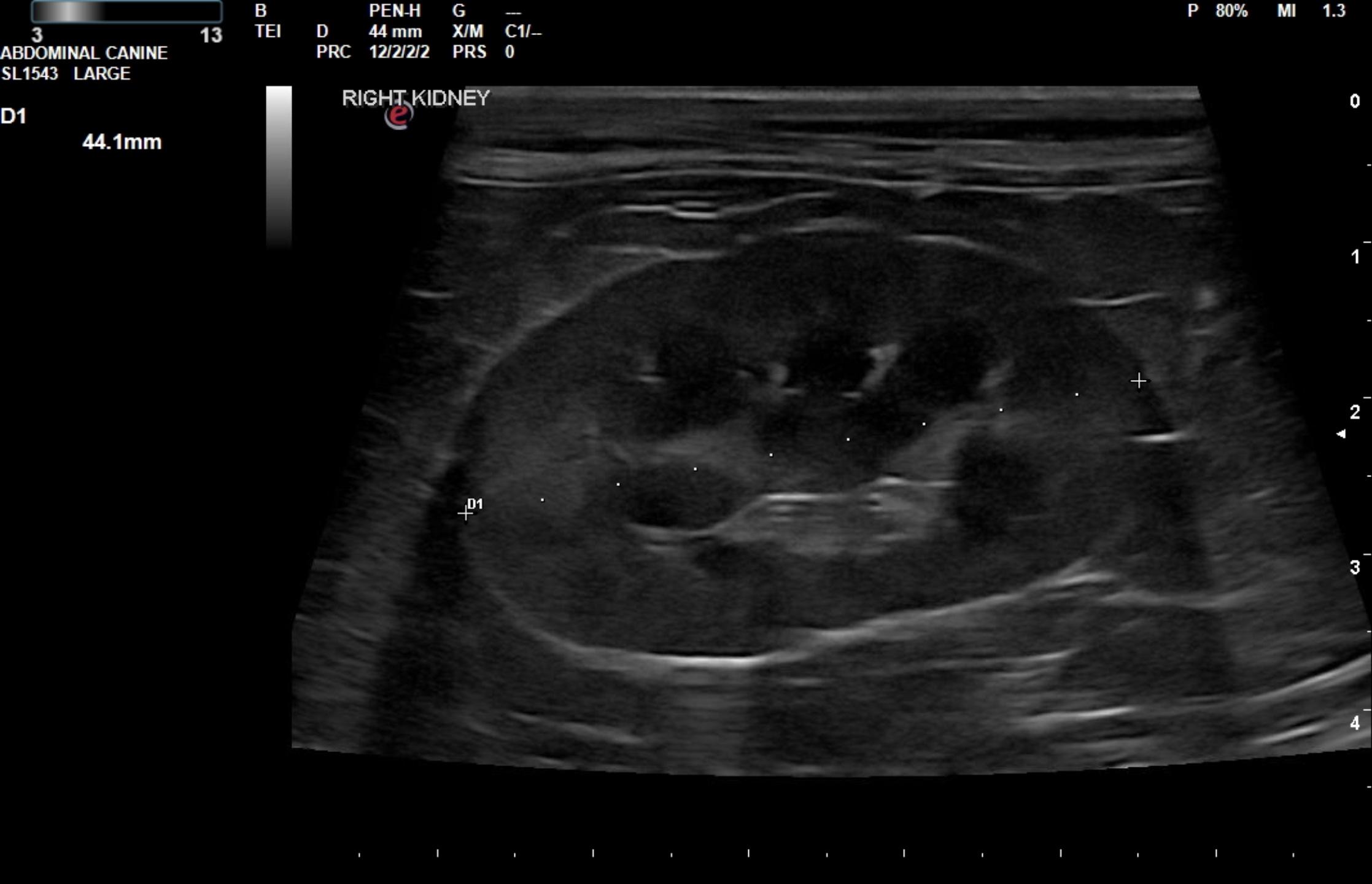
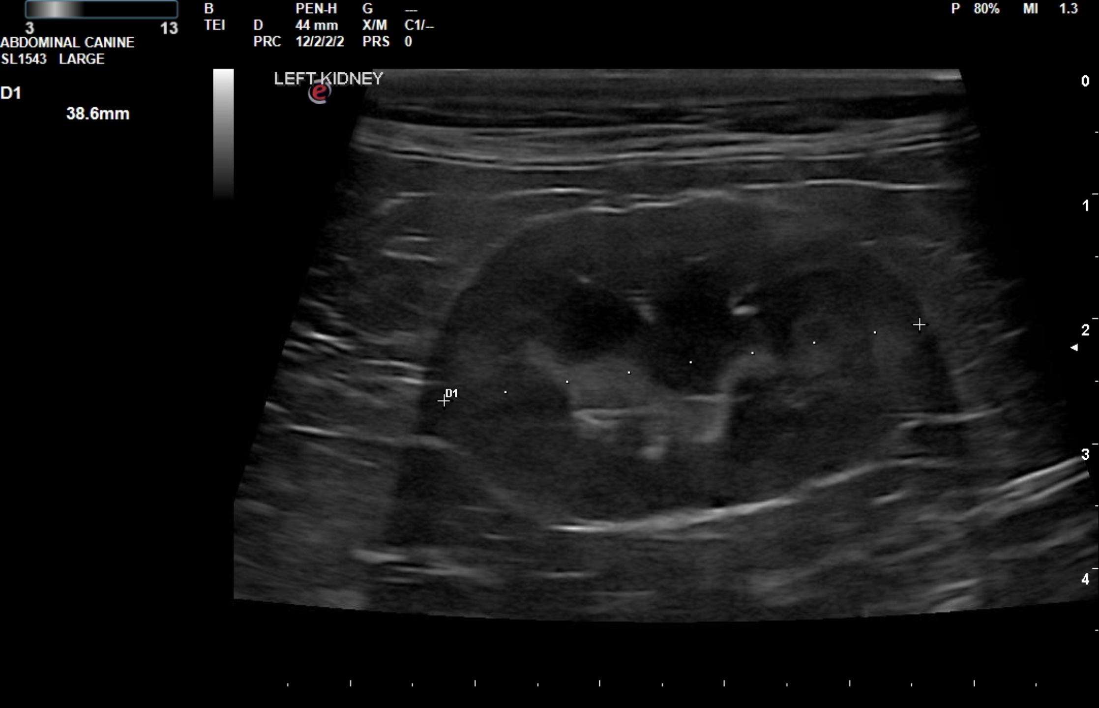
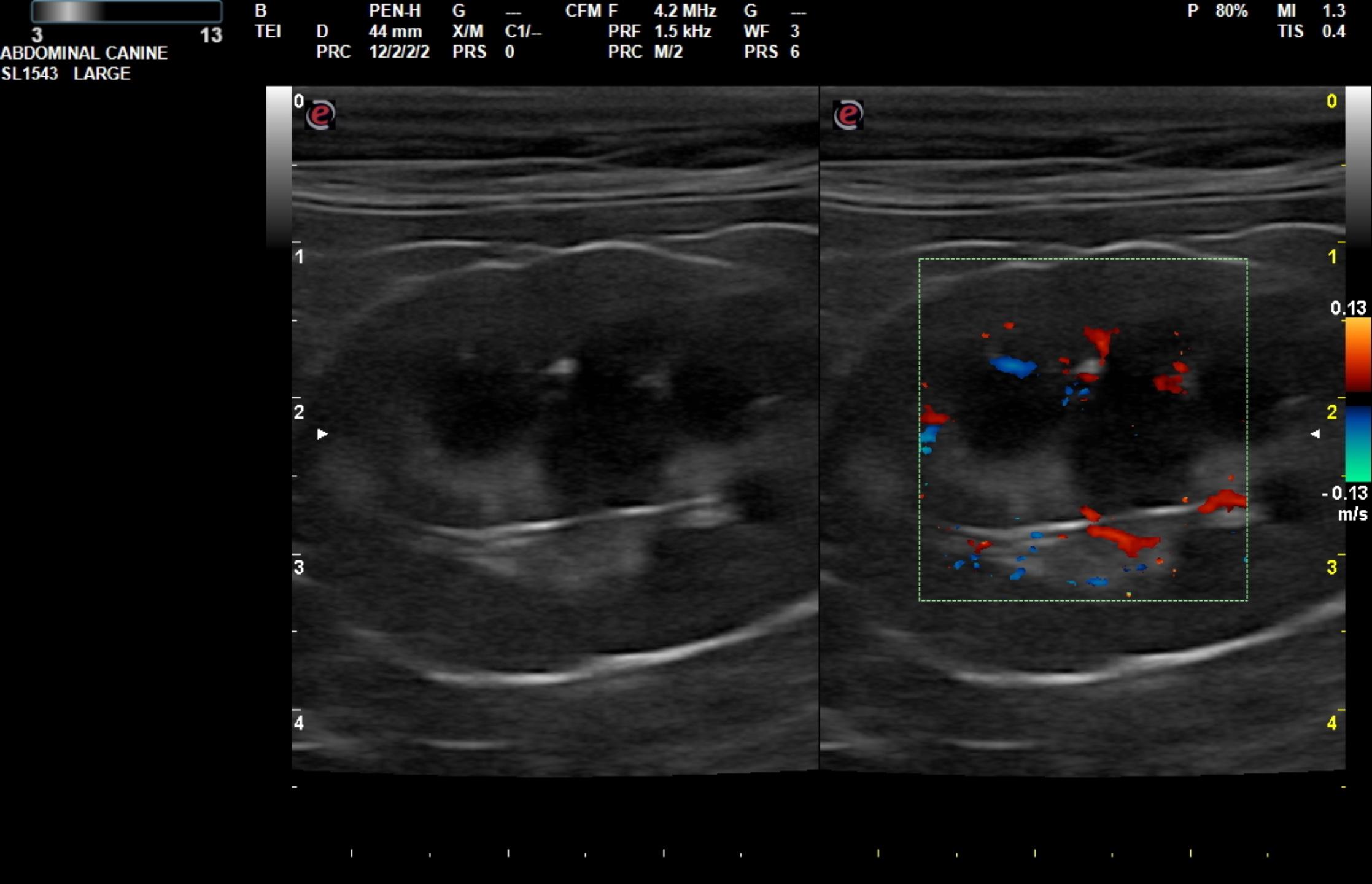
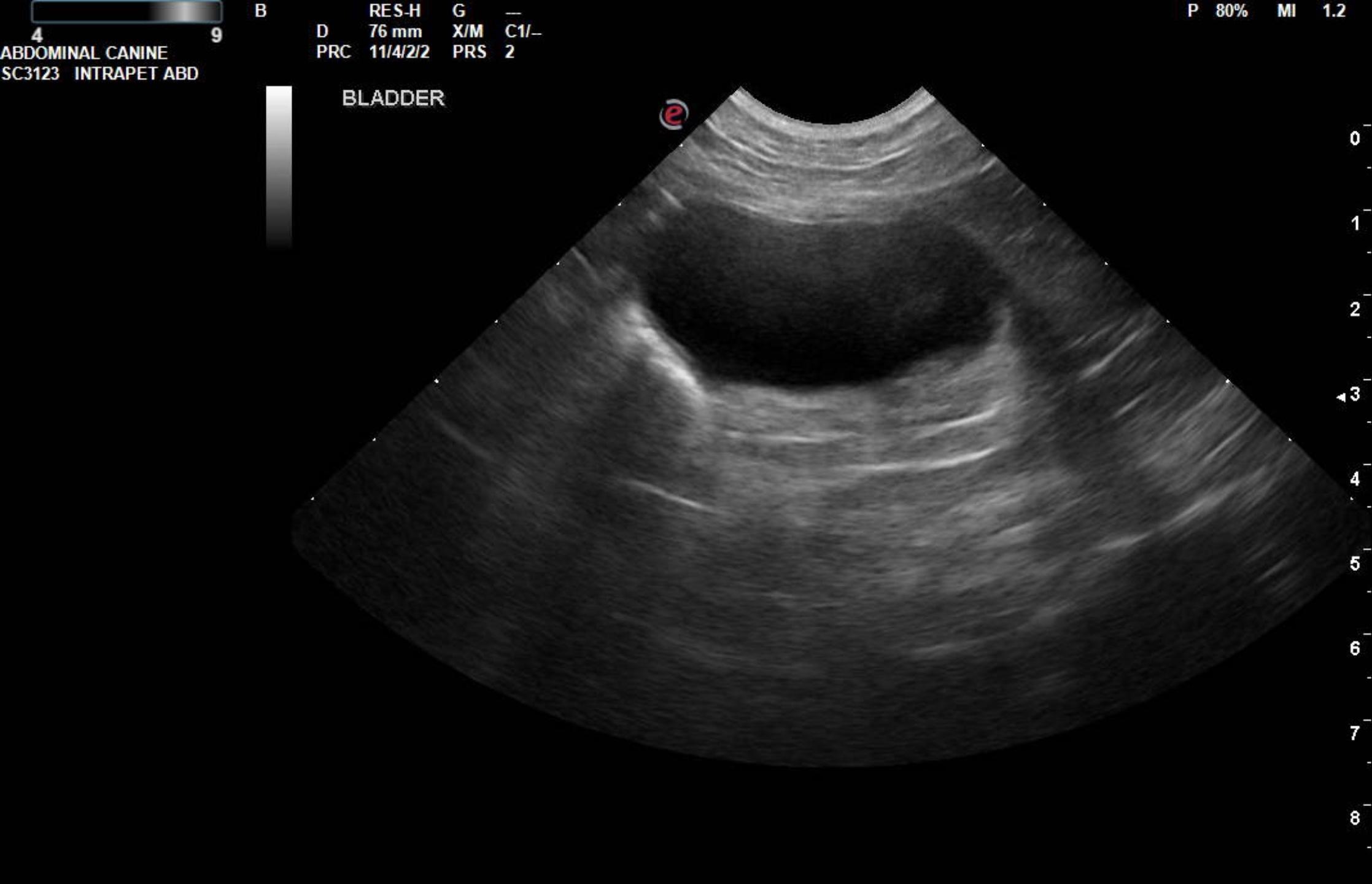
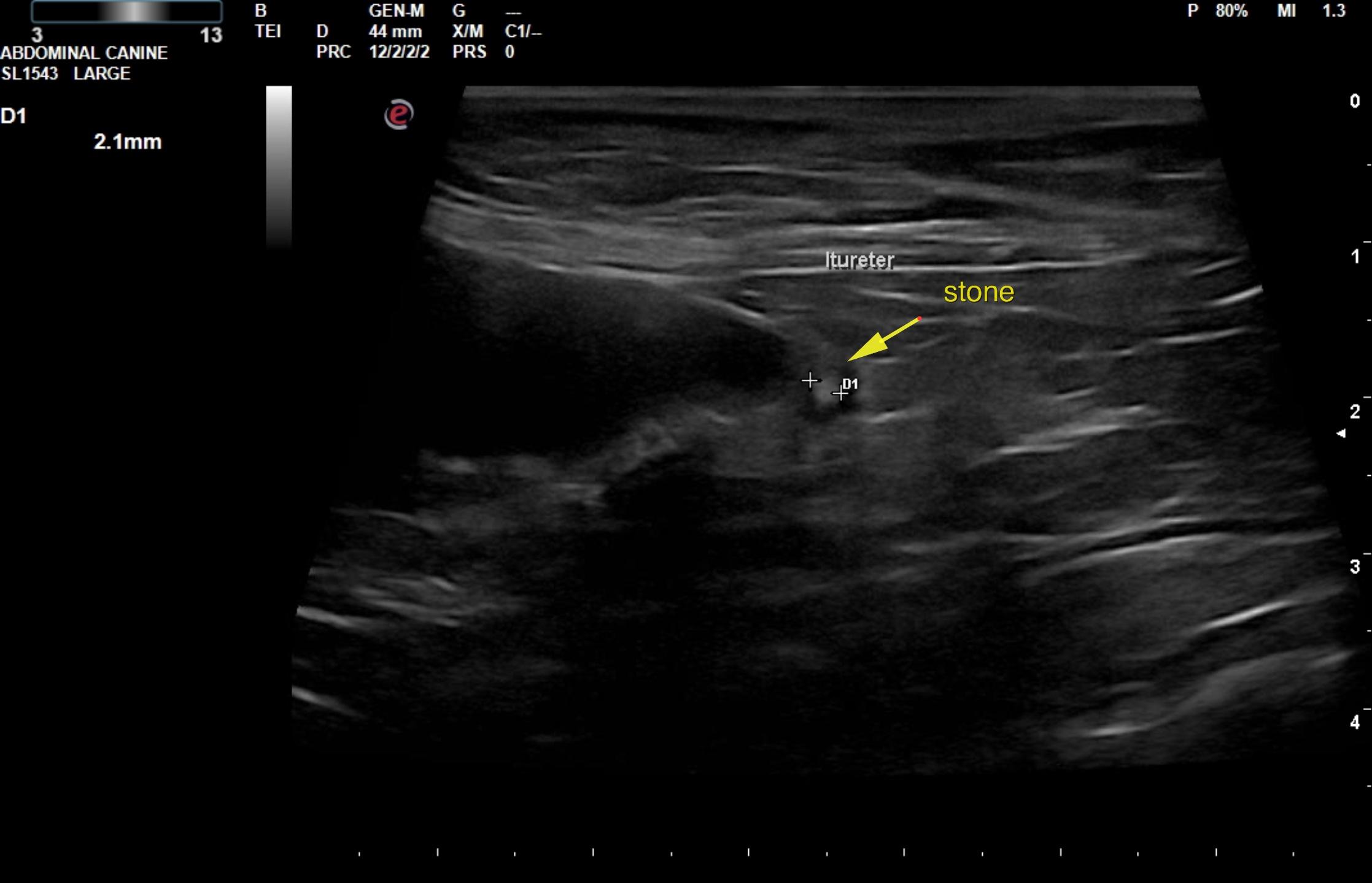
The urinary bladder itself was unremarkable. The pelvic urethra was imaged with no evident pathology. The right kidney measured 4.4 cm and was structurally unremarkable. Slight mineralization was noted at the corticomedullary junction. The left ureter in this patient revealed slight dilation with a focal, 0.21 cm calculus. This appears to be partially obstructive. It is small enough to potentially pass. Minor, hyperechoic inflammatory pattern was noted. Therefore, concurrent inflammation and irritation is likely an issue with this patient at the level of the ureteral papilla. Trace pyelectasia was noted in the left kidney. The left kidney measured 3.86 cm. Color flow Doppler assessment of the kidneys appeared to be adequate.
Otherwise, unremarkable abdomen. No bladder calculi at this current time. However,
the small calculus in the left ureter should pass given its size. Typically ureteral calculi
have been shown to pass if less than 0.3 cm with good ureteral function. This should be
monitored over the next 1-2 weeks to assess if this has potentially passed. Coverage
for urinary tract infection with double therapy such as Enrofloxacin and Cefa
combination may be best for this patient over the next 3-4 weeks. Caudal abdominal
focal palpation in the area of the cystourethral junction is recommended to assess for
clinical response to therapy.
Follow-up ultrasound 4 months later: The bladder revealed at least two small mobile calculi that appear to have dislodged from the ureteral papilla. However, remaining minor hydroureter was noted likely owing to stricture of the ureteral papilla owing to the stone history. The left ureter was persistently dilated and measured 0.2 cm similar to the prior sonogram. The remainder of the bladder was unremarkable. The kidneys revealed normal size and structure, corticomedullary definition and ratio for this age patient. The cortices presented largely uniform texture with normal echogenic relationship to liver and spleen. Medullary echogenicity differed distinctly from that of the cortex and no evidence or dilation could be seen. The capsules were acceptably uniform without dramatic irregularities. The left kidney measured 3.76 cm.mThe right kidney measured 4.32 cm.
Assessment: Obstructed left ureter. Two small mobile bladder calculi. They appear to have dislodged from the ureteral papilla. Unremarkable kidneys.