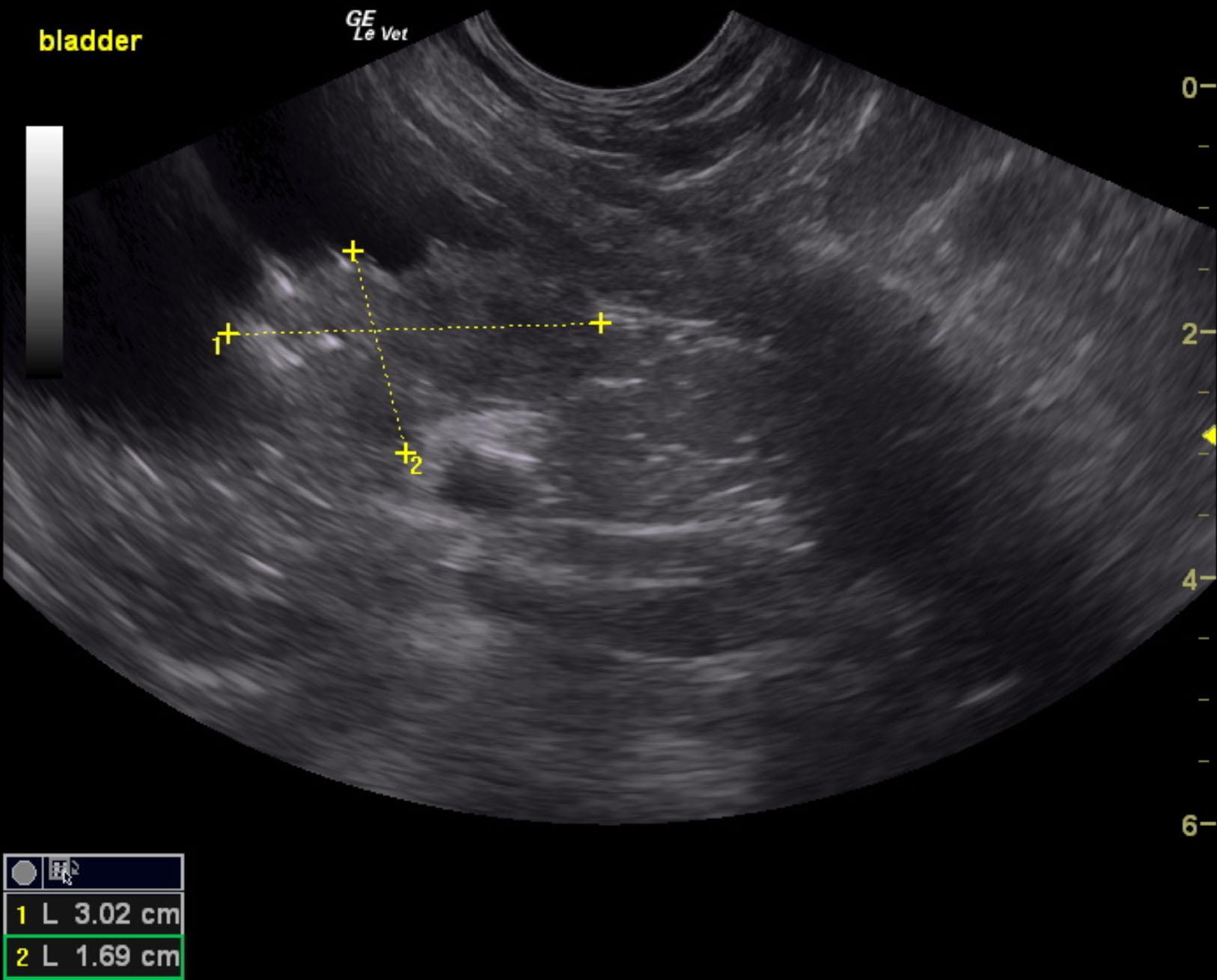
A 12-year-old FS Chow was presented for evaluation of persistent hematuria and stranguria. Urinalysis showed low-normal SG (1.020), hematuria, and leucosuria. Mildly elevated ALP activity was present on blood work.
A 12-year-old FS Chow was presented for evaluation of persistent hematuria and stranguria. Urinalysis showed low-normal SG (1.020), hematuria, and leucosuria. Mildly elevated ALP activity was present on blood work.