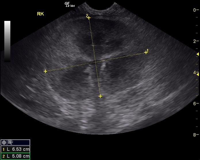
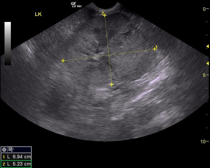
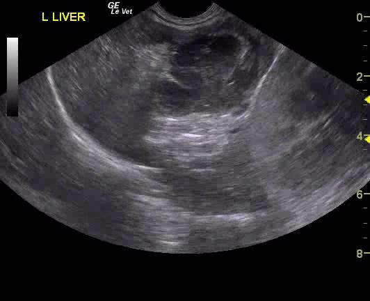
A 12½-year-old neutered male DSH cat with a history of hypertension controlled with amlodipine was presented for evaluation of PU/PD and weight loss. On physical examination, a cranial abdominal mass (suspected kidney) and moderate muscle atrophy was present. Abnormalities on CBC and serum biochemistry included neutrophilia and azotemia. Prior urine analysis had shown proteinuria with a SG of 1.028.
A 12½-year-old neutered male DSH cat with a history of hypertension controlled with amlodipine was presented for evaluation of PU/PD and weight loss. On physical examination, a cranial abdominal mass (suspected kidney) and moderate muscle atrophy was present. Abnormalities on CBC and serum biochemistry included neutrophilia and azotemia. Prior urine analysis had shown proteinuria with a SG of 1.028.