Subaortic stenosis (SAS) and pulmonic stenosis in a 11 year old MN German Shepherd dog

Case Study

Subaortic stenosis (SAS) and pulmonic stenosis in a 11 year old MN German Shepherd dog

An 11-month-old MN German Shepherd dog was presented for evaluation of a grade IV/VI heart murmur. Systemic BP was elevated (175/120). On survey radiographs cardiomegaly in the region of the right ventricle/pulmonary artery and mild left atrial enlargement was evident.
An 11-month-old MN German Shepherd dog was presented for evaluation of a grade IV/VI heart murmur. Systemic BP was elevated (175/120). On survey radiographs cardiomegaly in the region of the right ventricle/pulmonary artery and mild left atrial enlargement was evident.
Sonographic Differential Diagnosis

1.) Subaortic Stenosis – moderate to severe
2.) Pulmonic Stenosis – severe
3.) Ventricular arrhythmias

Image Interpretation

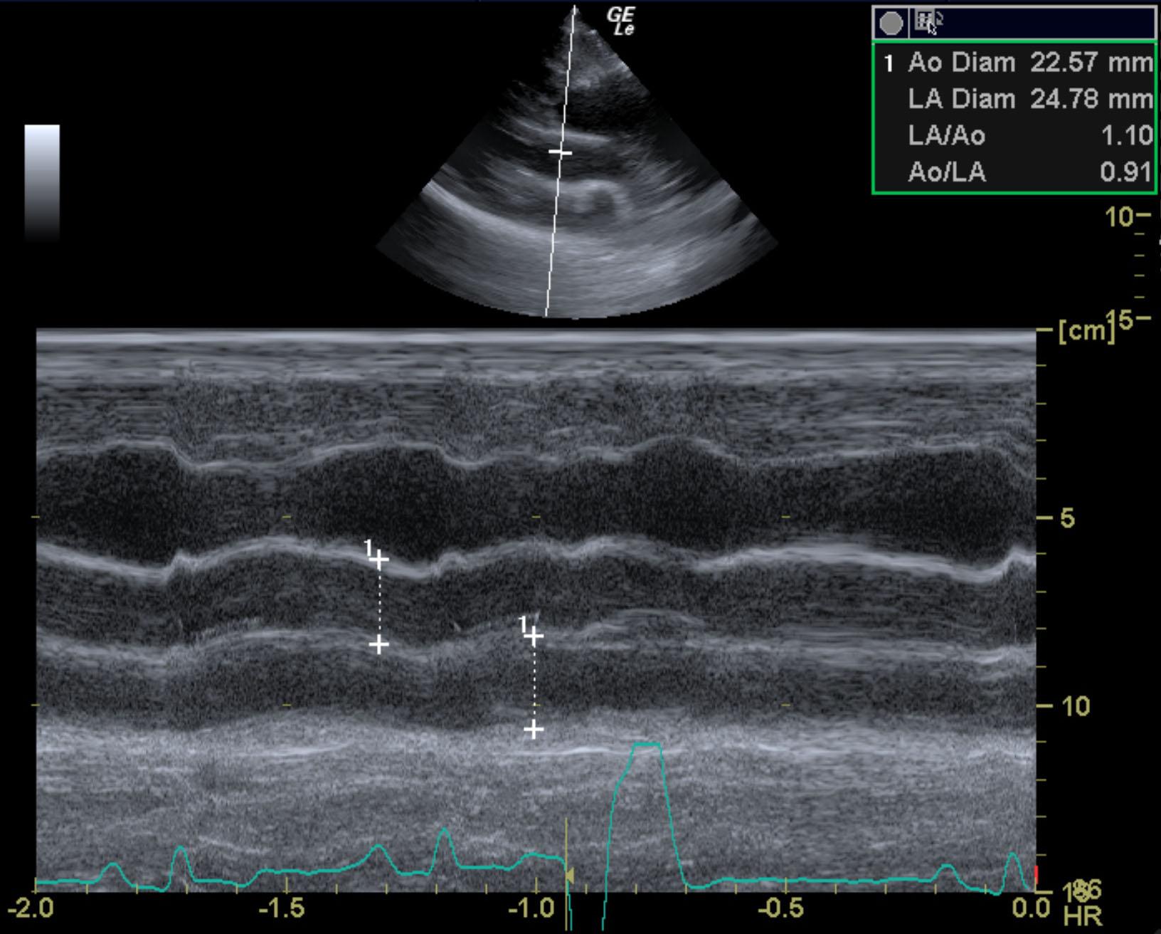
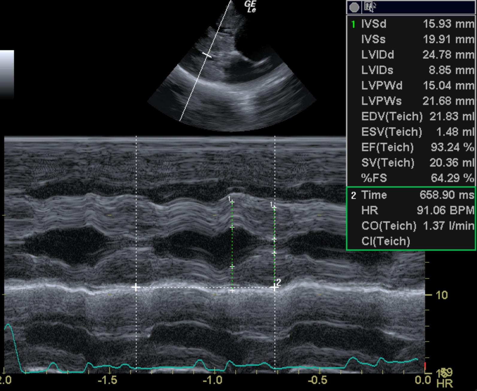
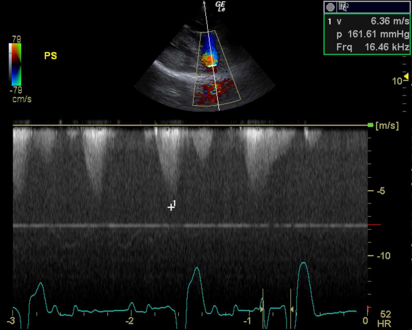
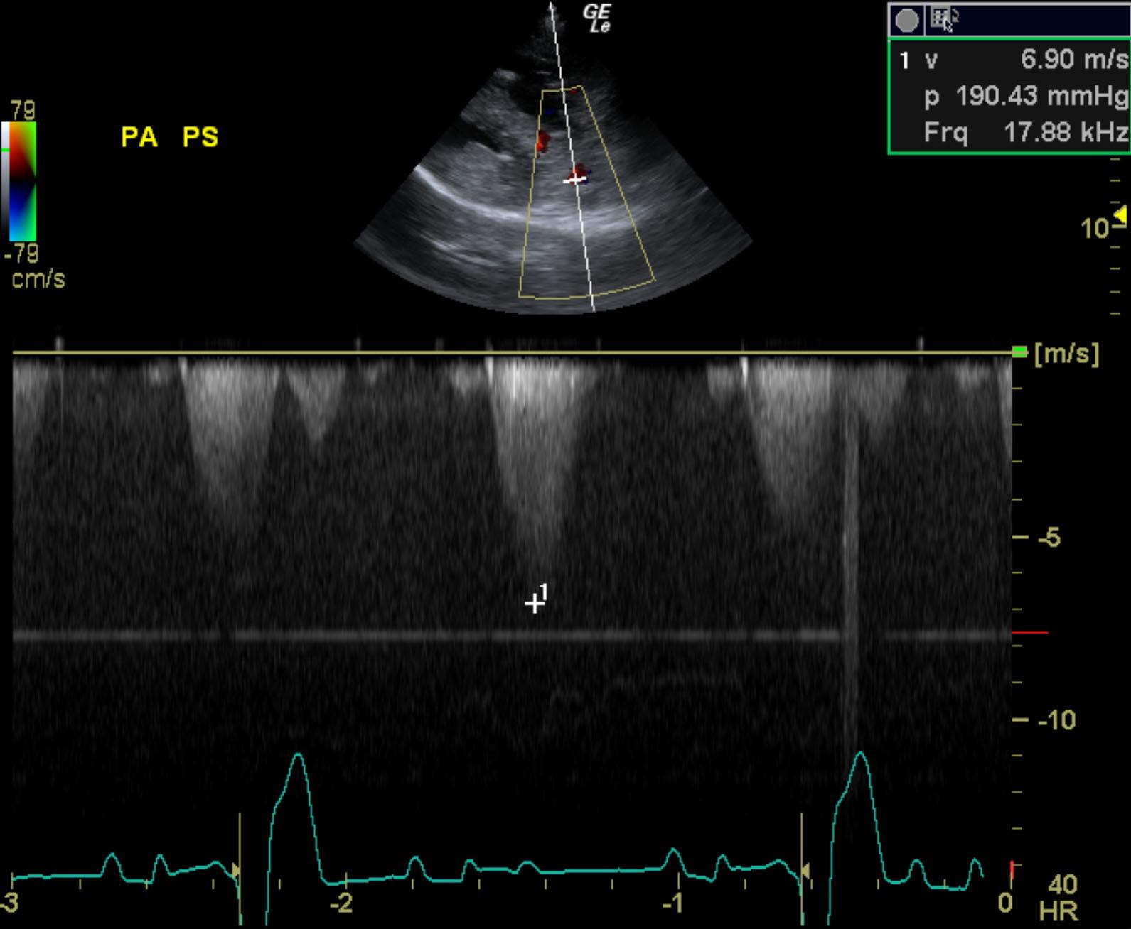
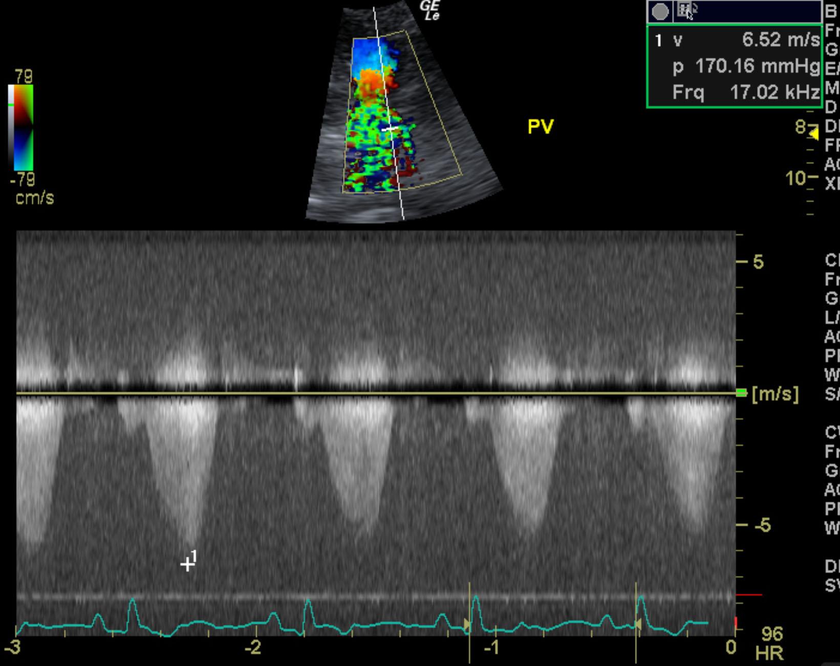
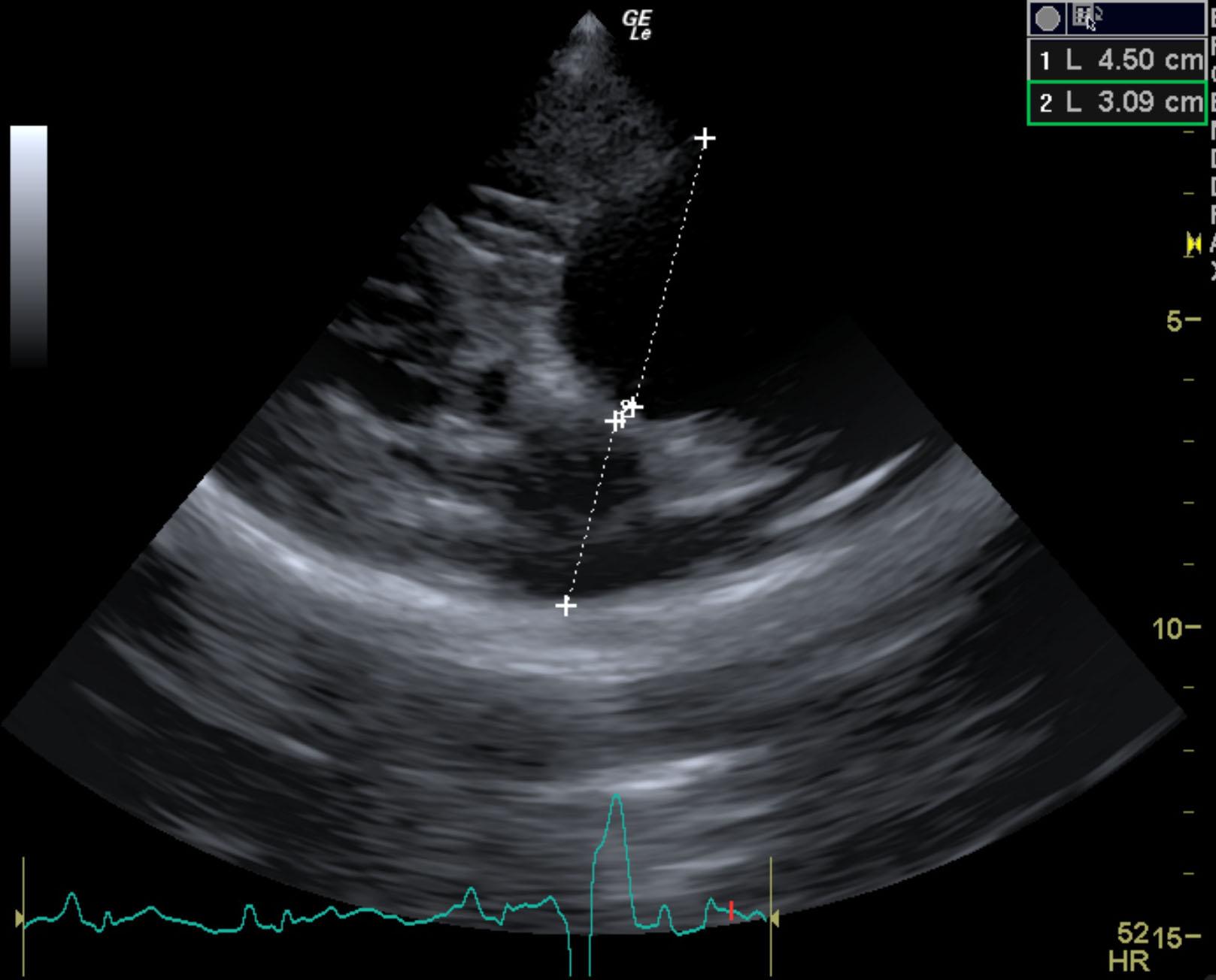
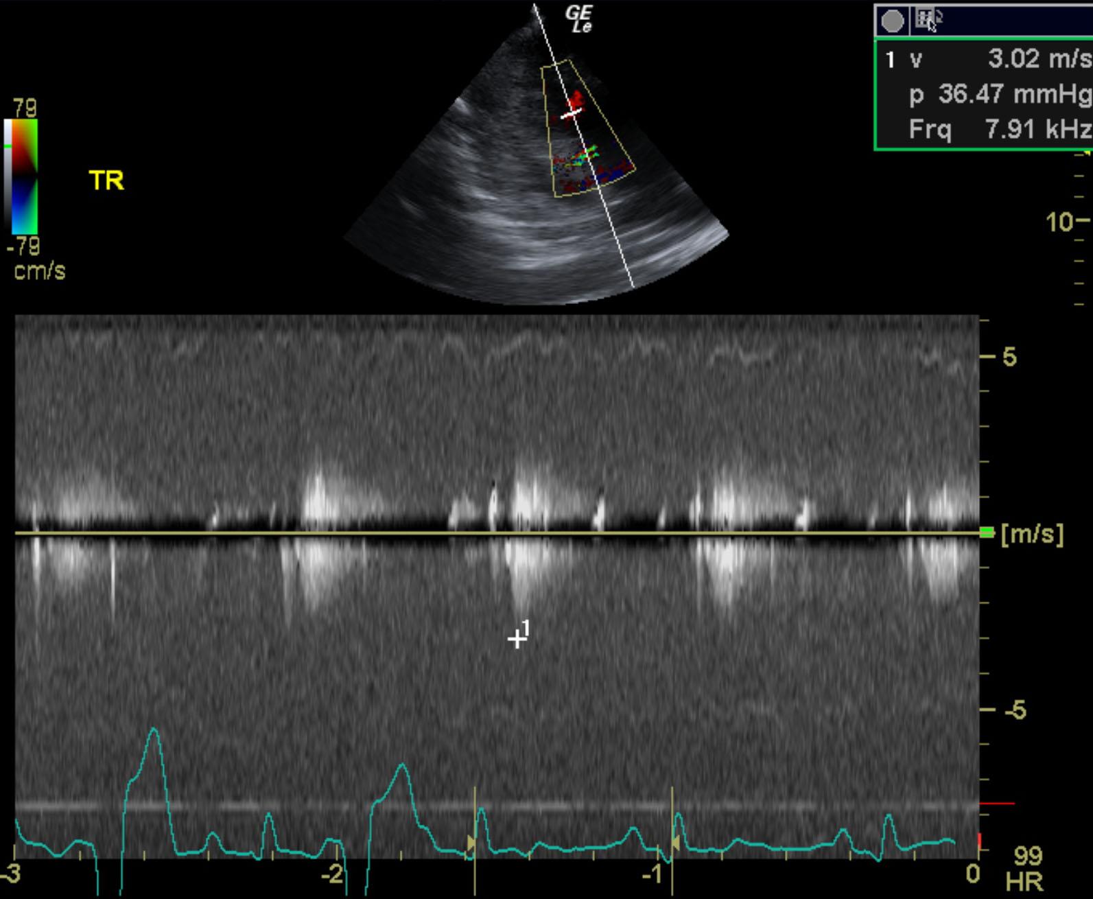
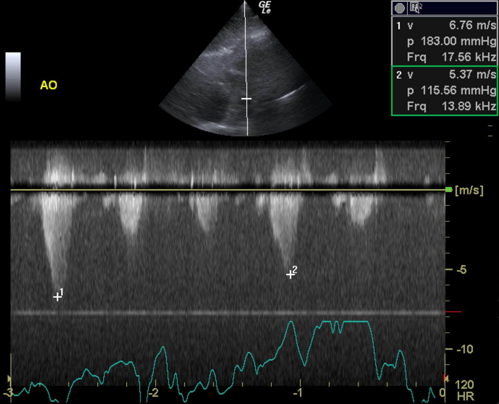
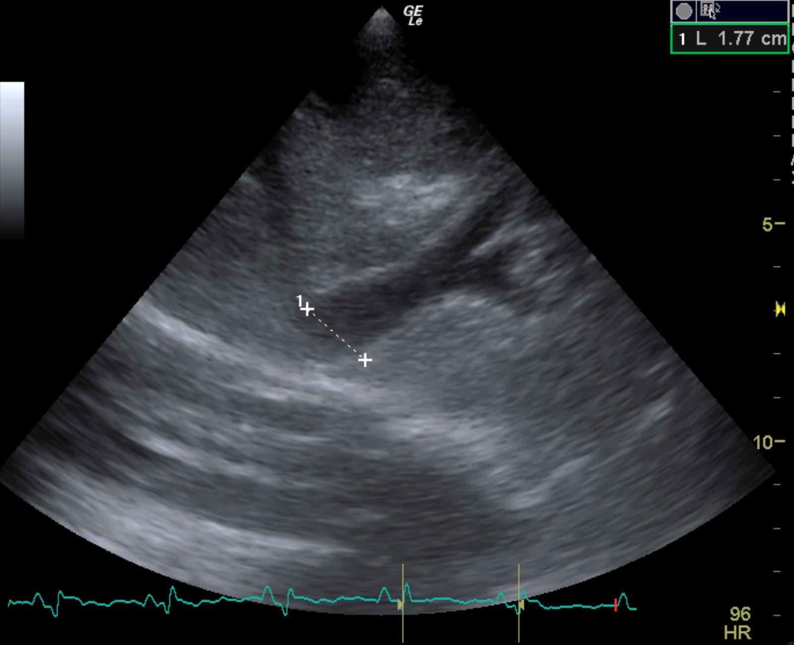
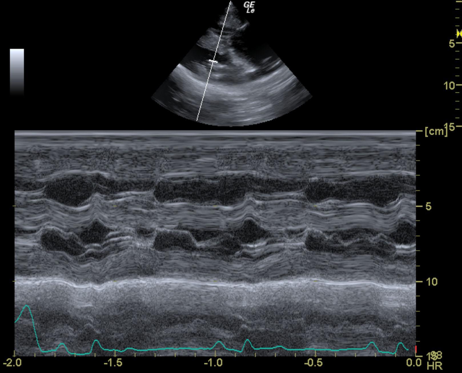
The left ventricle measures small in size, suggestive volume underload. There is adequate systolic function. There is mild to moderate LV concentric hypertrophy diffusely, subjectively and objectively based on MM measurements. The left atrium is normal in size (La:Ao 1.10) and the right atrium appears moderately to severely enlarged. The visible right heart is moderately to severely enlarged with severe concentric hypertrophy. The myocardium of the LV and RV is diffusely hyperechoic. Color Doppler identifies high velocity flow associated with the LVOT/AV and the PV. The peak velocity of aortic outflow is consistent with at least moderate SAS (LVOT 4.3-4.5m/s). The peak velocity of pulmonary outflow is consistent with severe PS (PV 6.9m/s). There is trivial AI, mild TR, and mild MR. No effusions or masses visualized. The caudal vena cava appears enlarged. There is a sinus rhythm with an average HR of 100-160bpm. There are single VPCs seen throughout the study.

DX

Subaortic Stenosis – moderate to severe, Pulmonic Stenosis – severe, Ventricular arrhythmias

Outcome

None

Clinical Differential Diagnosis

Congenital defect – pulmonic stenosis, aortic stenosis, ventricular septal defect

Sampling

None

Patient Information

Patient Name : Rookie L
Gender : Male, Neutered
Species : Canine
Type of Imaging : Ultrasound
Status : Complete
Liz Wuz Here : Yes
Code : 15-00208

Clinical Signs

  • Heart Murmur

Exam Finding

  • Heart Murmur

Images

rookie_langely_severe_ps_sas_gsd_11_mo_avelrookie_langely_severe_ps_sas_gsd_11_mo_cvcrookie_langely_severe_ps_sas_gsd_11_mo_epssrookie_langely_severe_ps_sas_gsd_11_mo_la_aorookie_langely_severe_ps_sas_gsd_11_mo_lv_mmoderookie_langely_severe_ps_sas_gsd_11_mo_ps_cf_2rookie_langely_severe_ps_sas_gsd_11_mo_ps_cf_3rookie_langely_severe_ps_sas_gsd_11_mo_ps_cfrookie_langely_severe_ps_sas_gsd_11_mo_ra_larookie_langely_severe_ps_sas_gsd_11_mo_tr

Clinical Signs

  • Heart Murmur