The patient is a canine Shih Tzu, NM, 13 years. The patient has been coughing and has a grade III/VI heart murmur.
The patient is a canine Shih Tzu, NM, 13 years. The patient has been coughing and has a grade III/VI heart murmur.
Case Study
Stage B1 Valvular Disease, Mitral valve prolapse. Slight left atrial enlargement in a MN 13 year old Shih Tzu Dog
Sonographic Differential Diagnosis
Mitral valve prolapse. Slight left atrial enlargement.
Primary respiratory protocol is warranted and assessment for tracheal collapse or primary respiratory disease.
Image Interpretation
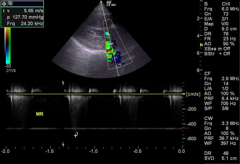
The left atrium was slightly prominent, yet not to the point of causing congestive heart failure or respiratory signs. No evidence of “smoke” or thrombotic activity was noted. The atrial septum was straight without evidence of dilation or contraction. The LA/AO ratio was within normal limits. The cranial and caudal mitral valve leaflets demonstrated mildly vegetative contour. Prolapse of the anterior leaflet was noted. The left ventricle demonstrated normal volume, septal and free wall diastolic thicknesses, and papillary integrity without evidence of significant dilation or contraction. Ventricular function was deemed adequate expressed by the fractional shortening measurement listed below. The aortic outflow tract revealed direct laminar flow along the ventricular septum with normal outflow velocity through linear valve leaflets with adequate extension, closure and uninterrupted motion. No evidence was found regarding valvular insufficiency or post stenotic dilation. Periaortic tissue was uniform and of normal echogenicity. No inconsistencies were revealed regarding the presence of an aortic body tumor or chemodectoma. The right atrium was subjectively assessed and found to be uniformly smaller than the left atrium without evidence of dilation or contraction. No neoplastic evidence was visualized here or into its extension of the right auricle. The tricuspid valve was found to be linear with proper extension, length and closure. The right ventricle demonstrated normal size (1/3 diameter of the left ventricle), morphology, and kinetic activity. No evidence of heartworm or infiltrative disease was noted. The pulmonic outflow tract presented free of dilation (1:1 PA/AO ratio), adequate laminar flow, no evidence of insufficiency, and free of visible heartworm activity. Variable areas of lung consolidation and comet tail pattern was noted in the lungs. This is suggestive for thromboembolic disease, subacute respiratory distress syndrome, pneumonitis, or non cardiogenic pulmonary edema. Mitral insufficiency velocity 5.8 m/sec.
Tricuspid insufficiency velocity 1.9 m/sec.
DX
Stage B1 Valvular Disease. Mitral valve prolapse. Slight left atrial enlargement.
Outcome
None
Sampling
None
Video
Patient Information
Patient Name :
Tobey S
Gender :
Male, Neutered
Species :
Canine
Type of Imaging : Ultrasound
Status :
Complete
Liz Wuz Here :
Yes
Code :
15-00158
Clinical Signs
- Coughing
- Heart Murmur
Exam Finding
- Heart Murmur
Images
Clinical Signs
- Coughing
- Heart Murmur