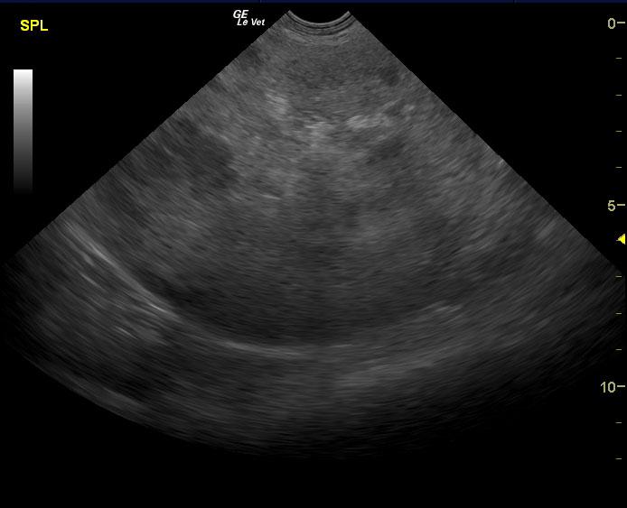
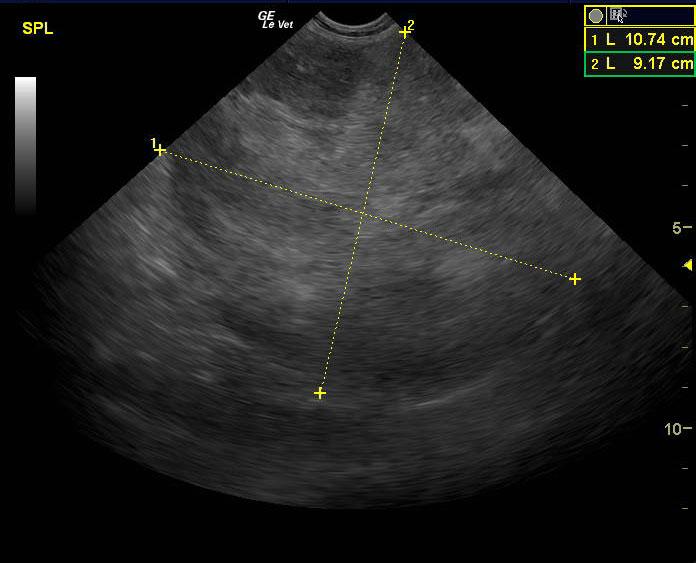
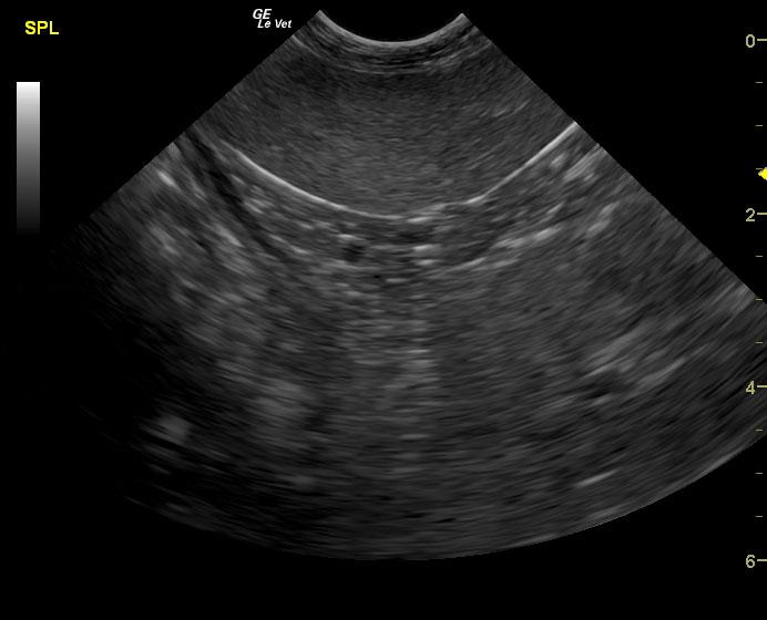
A 14-year-old SF Chow mix was presented for evaluation of weakness, which had also been present a week ago. On physical examination, a palpable abdominal mass was present. Severe non-regenerative anemia and hypoalbuminemia were present on CBC and blood chemistry.
A 14-year-old SF Chow mix was presented for evaluation of weakness, which had also been present a week ago. On physical examination, a palpable abdominal mass was present. Severe non-regenerative anemia and hypoalbuminemia were present on CBC and blood chemistry.