The patient is a neutered male, 12 year old, Havanese dog. There is concern for enlarged spleen on abdominal palpation. Bloodwork abnormalities include albumin of 2.3 and ALP of 357. The dog is also anemic with an HCT of 23%
The patient is a neutered male, 12 year old, Havanese dog. There is concern for enlarged spleen on abdominal palpation. Bloodwork abnormalities include albumin of 2.3 and ALP of 357. The dog is also anemic with an HCT of 23%
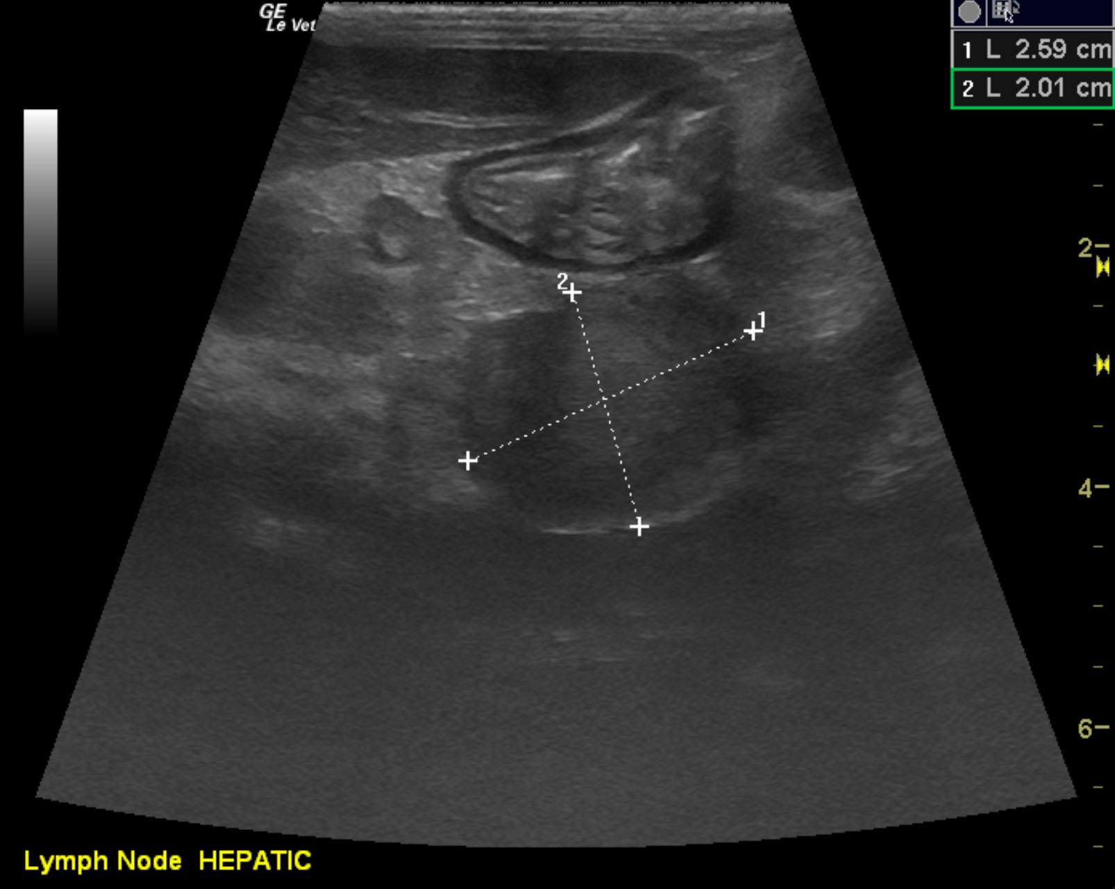
Given the hepatic lymphadenopathy, infiltrative disease is likely. Multicentric lymphoma pattern or similar neoplasia.
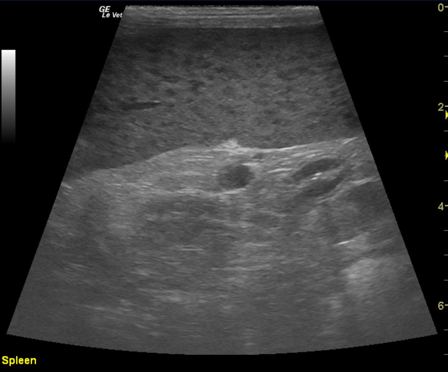
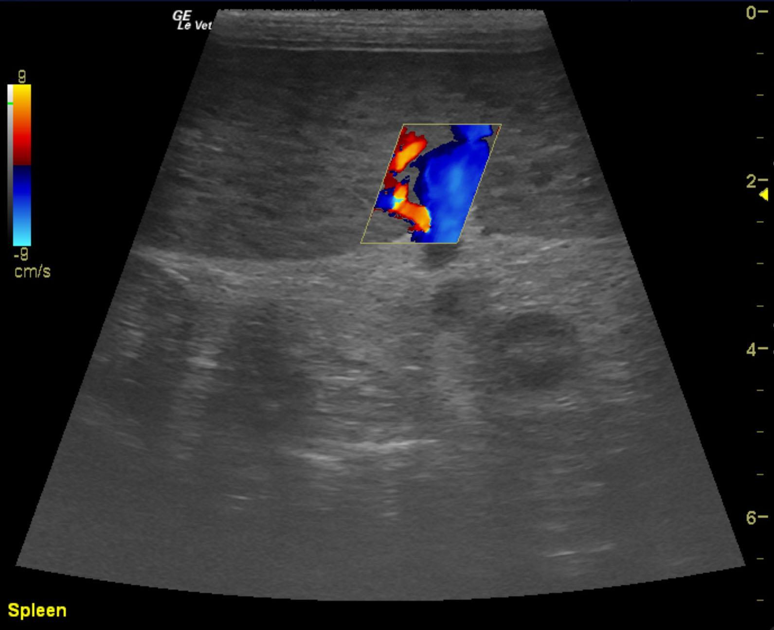
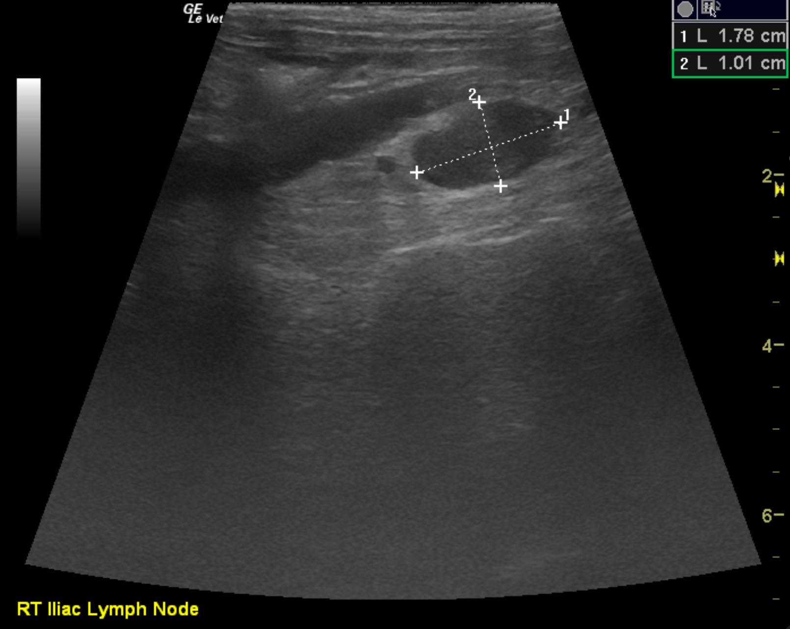
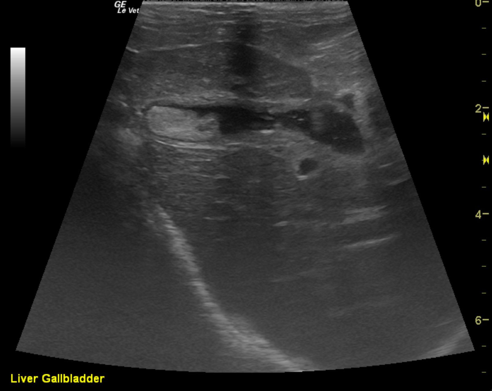
The spleen was enlarged with micronodular changes and a scalloping contour. The liver was swollen and mildly irregular. A hepatic lymph node was mildly enlarged and measured 1.13 x 0.9 cm. A separate, larger node was noted and measured 2.6 x 2.01 cm. The inguinal lymph nodes were enlarged, hypoechoic and irregular. Iliac lymph nodes were also enlarged and measured 1.8 cm. Regional inflammatory pattern was noted.
None
Cranial mediastinal imaging and chest radiographs are recommended.
Splenic neoplasia, GI/other neoplasia, GI blood loss/gastroenteritis, protein-losing nephropathy, bone marrow disease.
Ultrasound-guided FNA of the spleen and hepatic lymph nodes were completed without complication. Cytology revealed lymphoma.