A 7-year-old SF PBGV was presented for evaluation of a grade III/VI systolic murmur.
A 7-year-old SF PBGV was presented for evaluation of a grade III/VI systolic murmur.
Case Study
Severe Pulmonic Stenosis, Right Ventricular Hypertrophy, Right Atrial Enlargement in a PBGV female dog.
Sonographic Differential Diagnosis
Pulmonic stenosis with mild to moderate concentric right ventricular hypertrophy and right atrial enlargement. Recommend a referral to an interventional cardiologist for potential balloon valvuloplasty especially given the elevated heart rate in this patient.
Image Interpretation
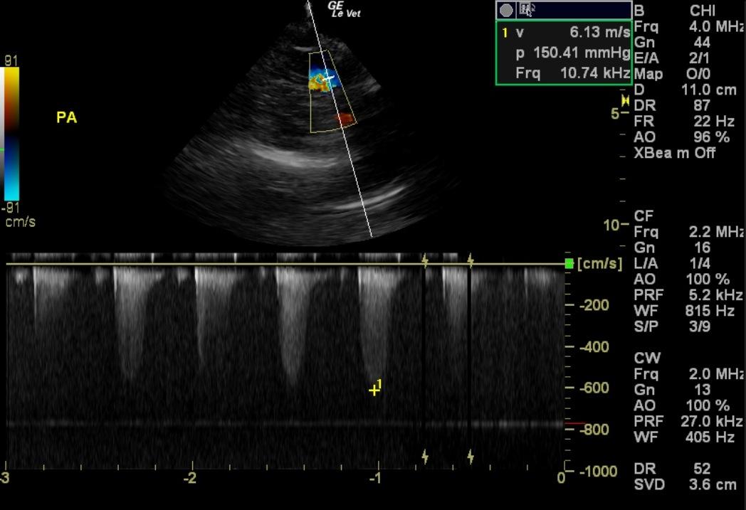
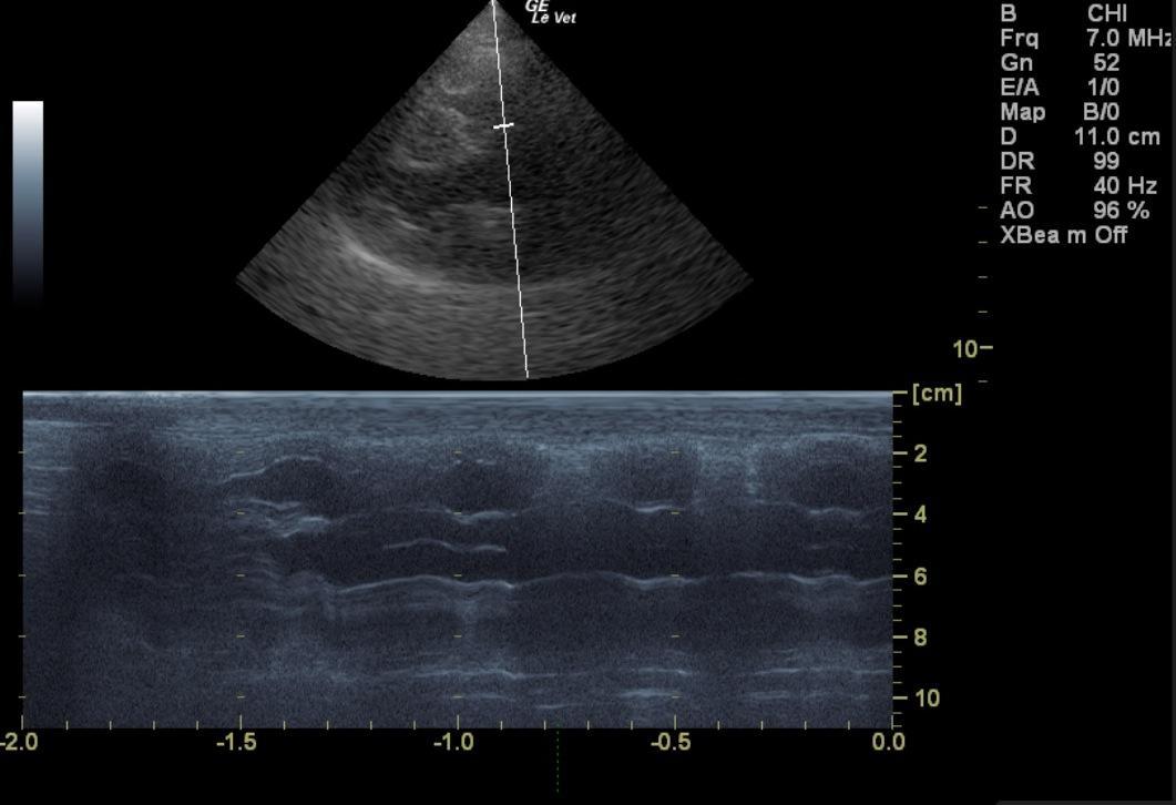
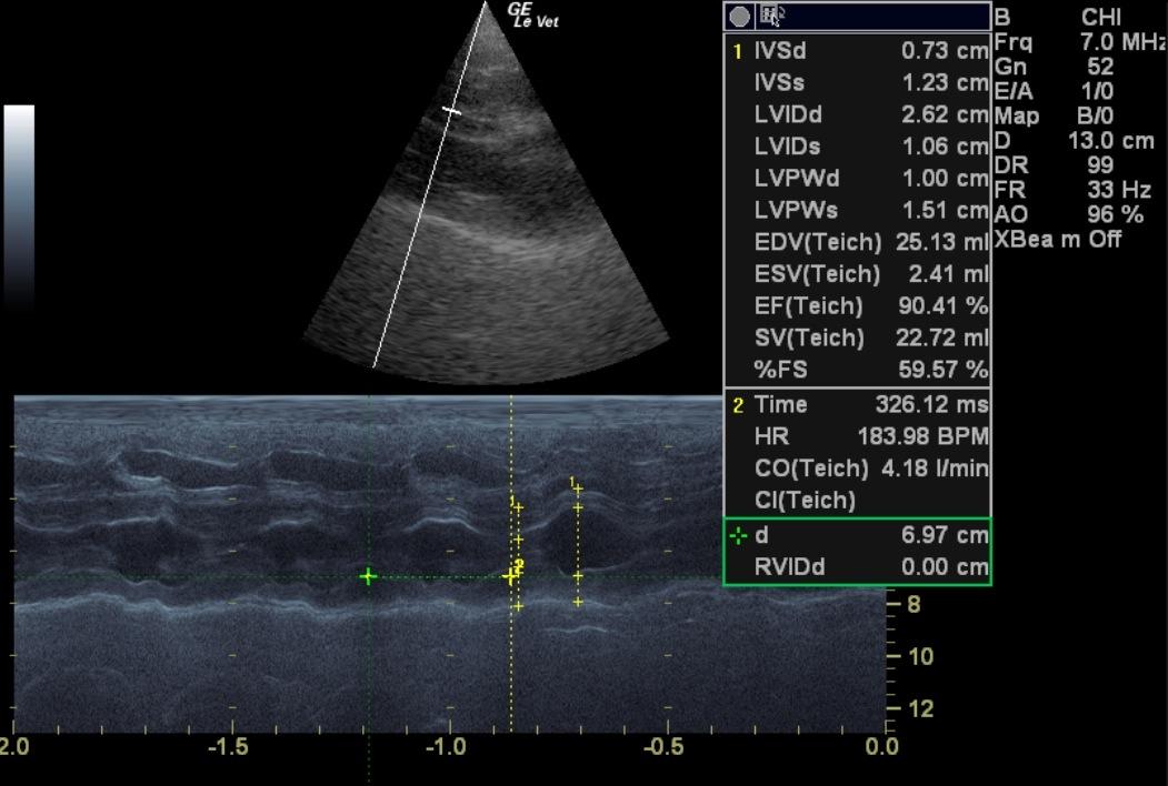
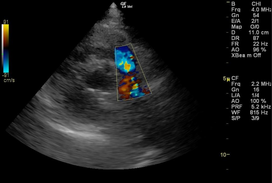
The echocardiogram in this patient demonstrated normal left atrial size and structure with no evidence of “smoke” or thrombi. The cranial and caudal mitral valve leaflets presented normal linear structure, extension in systole, and union in diastole. No significant dystrophic or vegetative changes were noted. The left ventricle presented normal free wall and septal thicknesses with linear contour. The chamber was neither dilated nor restricted. The myocardium presented normal echogenicity without evidence of significant fibrotic or ischemic disease. Contractility of the ventricular walls was adequate and in normal range for this breed and patient size. The left ventricular outflow tract demonstrated normal laminar flow without evidence of hemodynamically significant insufficiency or excessive outflow velocity. Tricuspid insufficiency was noted with mild to moderate right atrial enlargement. The right ventricle presented concentric hypertrophy. The pulmonary outflow tract revealed a thickened, pulmonic valve with dysplastic changes and severe elevations in velocity at 6.1 m/sec. Pulmonic insufficiency was noted and likely secondary to the primary pulmonic stenosis. No visible pericardial or free pleural fluid was noted. No echographically detectable evidence of infiltrative disease was visible. The mediastinum was free of masses in the visible window. The diaphragm was visualized without interruption. Tricuspid insufficiency 2.1 m/sec.
DX
Pulmonic stenosis with mild to moderate concentric right ventricular hypertrophy and right atrial enlargement.
Outcome
No further outcome at this time.
Clinical Differential Diagnosis
Mitral/tricuspid endocardiosis Vegetative endocarditis Cardiomyopathy Progressive congenital defect – aortic/tricuspid stenosis, tricuspid dysplasia Anemia/hyperviscosity Hypertension
Sampling
None
Video
Patient Information
Patient Name :
Maggie S
Gender :
Female, Spayed
Species :
Canine
Type of Imaging : Ultrasound
Status :
Complete
Liz Wuz Here :
Yes
Code :
15-00144
Clinical Signs
- Heart Murmur
Images
Clinical Signs
- Heart Murmur