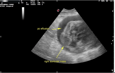
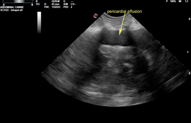
A 12-year-old MN Dachshund was presented for evaluation of abdominal distension that had been noticed after grooming. Additional history was that after grooming anorexia and vomiting had developed. Abnormalities CBC and serum biochemistry were leukocytosis and mildly elevated ALT activity (239). Survey radiographs showed cardiomegaly, pulmonary edema, pleural effusion, and splenomegaly.
A 12-year-old MN Dachshund was presented for evaluation of abdominal distension that had been noticed after grooming. Additional history was that after grooming anorexia and vomiting had developed. Abnormalities CBC and serum biochemistry were leukocytosis and mildly elevated ALT activity (239). Survey radiographs showed cardiomegaly, pulmonary edema, pleural effusion, and splenomegaly.