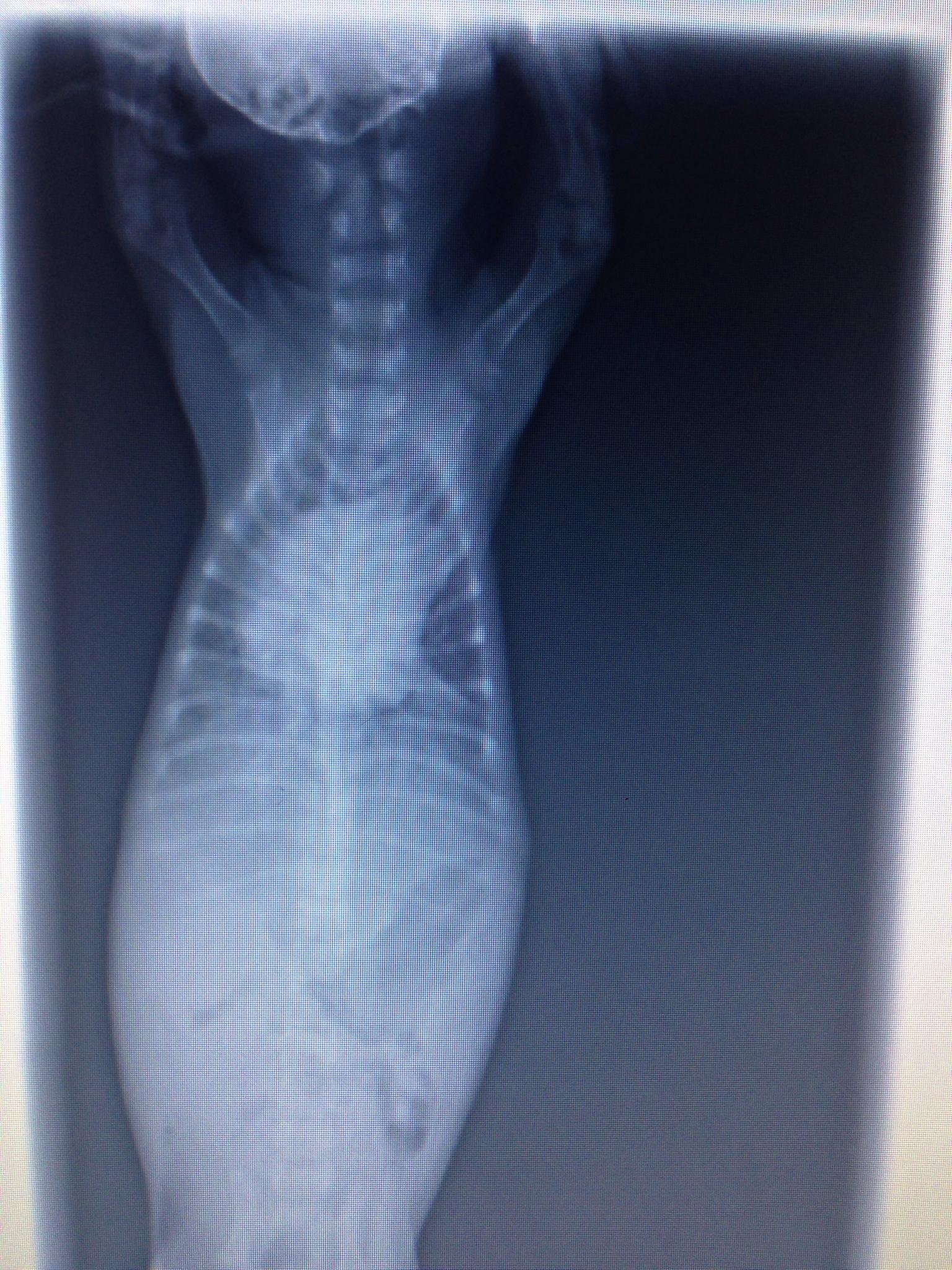
An 8-week-old female Silky Terrier was presented for evaluation of dyspnea and diarrhea. No murmur was auscultated on physical examination. Parvo snap test was negative and hematocrit was 28%. Survey radiographs show right-sided cardiomegaly, focal area of pulmonary consolidation, and an enlarged PA.
An 8-week-old female Silky Terrier was presented for evaluation of dyspnea and diarrhea. No murmur was auscultated on physical examination. Parvo snap test was negative and hematocrit was 28%. Survey radiographs show right-sided cardiomegaly, focal area of pulmonary consolidation, and an enlarged PA.