Case Study
RADS – Peritonitis with possible Foreign Body in a 10 year old MN Dachshund dog
Image Interpretation
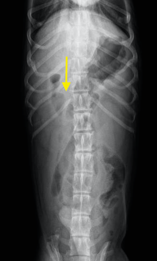
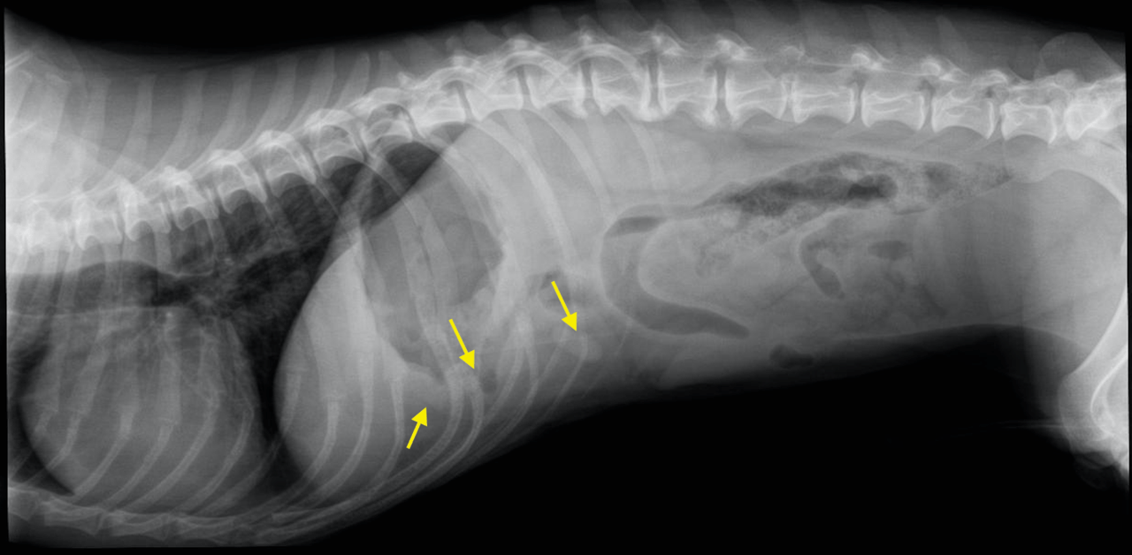
right lateral and VD abdomen – Osseous structures:
Moderate degenerative changes were associated with the axial skeleton empahsized in the thoracolumbar area including moderate Spondylosis deformans, vertebral endplate sclerosis, mild and moderate intervertebral disc space collapse L1/2 and L3/4 respectively as well as multifocal intervertebral disc mineralization. The opacity oft he small synovial joints was decreased at L3/4 compatible with prior hemilaminectomy.
Intraabdominal structures:
The serosal detail was mildly reduced emphasizing the cranial abdomen caudal to the stomach. The pylorus presented moderate caudal and ventral displacement. The wall oft he gastric corpus and pyloric antrum appeared to be thickened.
The liver was hardly assessable but appeared to be of normal size.
The spleen and kidneys were within normal limits.
The urinary bladder was in a relatively caudal position. The prostate was not seen.
There was no sign of complete mechanical intestinal obstruction but a well delineated ovoid radioopaque structure was associated with the intestinal loops (arrowed) on the lateral view.
Intrathoracic structures:
There was hypovolemia with underperfusion of the lung. The cardiac silhouette was within normal limits for size and shape. The lung parenchyma was within normal limits as far as included.
DX
Comments
|
Possible underlying causes include inflammatory – or less likely neoplastic – infiltration of the pancreas, stomach, liver & gallbladder or a combination of the aforementioned.
|
Patient Information
Images