RADS – Non-Cardiogenic Pulmonary Edema, Chronic Bronchial Pattern and Pulmonary Nodule in a 10 year old MN Siberian Husky dog

Case Study

RADS – Non-Cardiogenic Pulmonary Edema, Chronic Bronchial Pattern and Pulmonary Nodule in a 10 year old MN Siberian Husky dog

Presented for first episode of seizure activity earlier today, lasting about 30 seconds. Physical exam WNL. Chemistry unremarkable other than slightly elevated glucose. 

Presented for first episode of seizure activity earlier today, lasting about 30 seconds. Physical exam WNL. Chemistry unremarkable other than slightly elevated glucose. 

DX

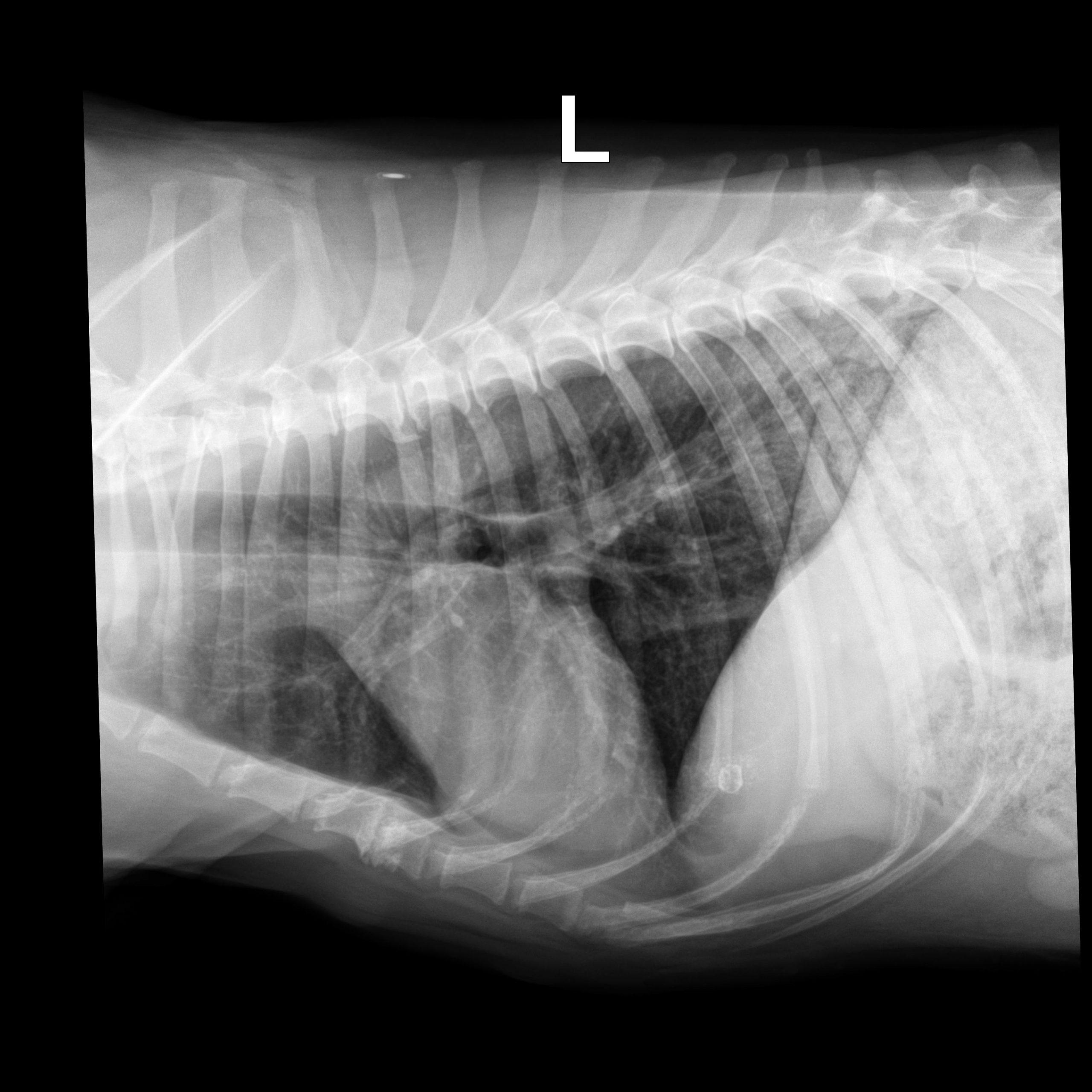
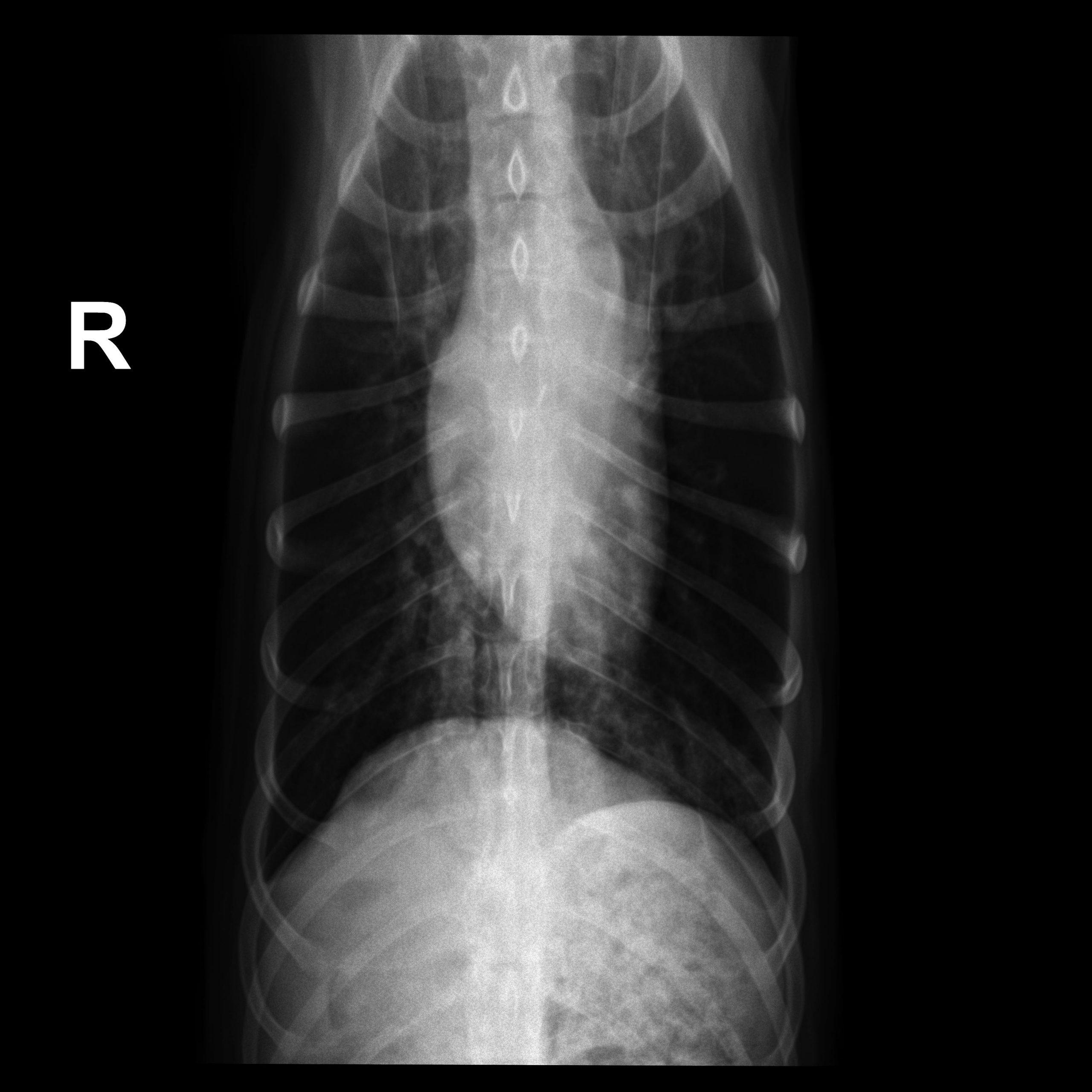
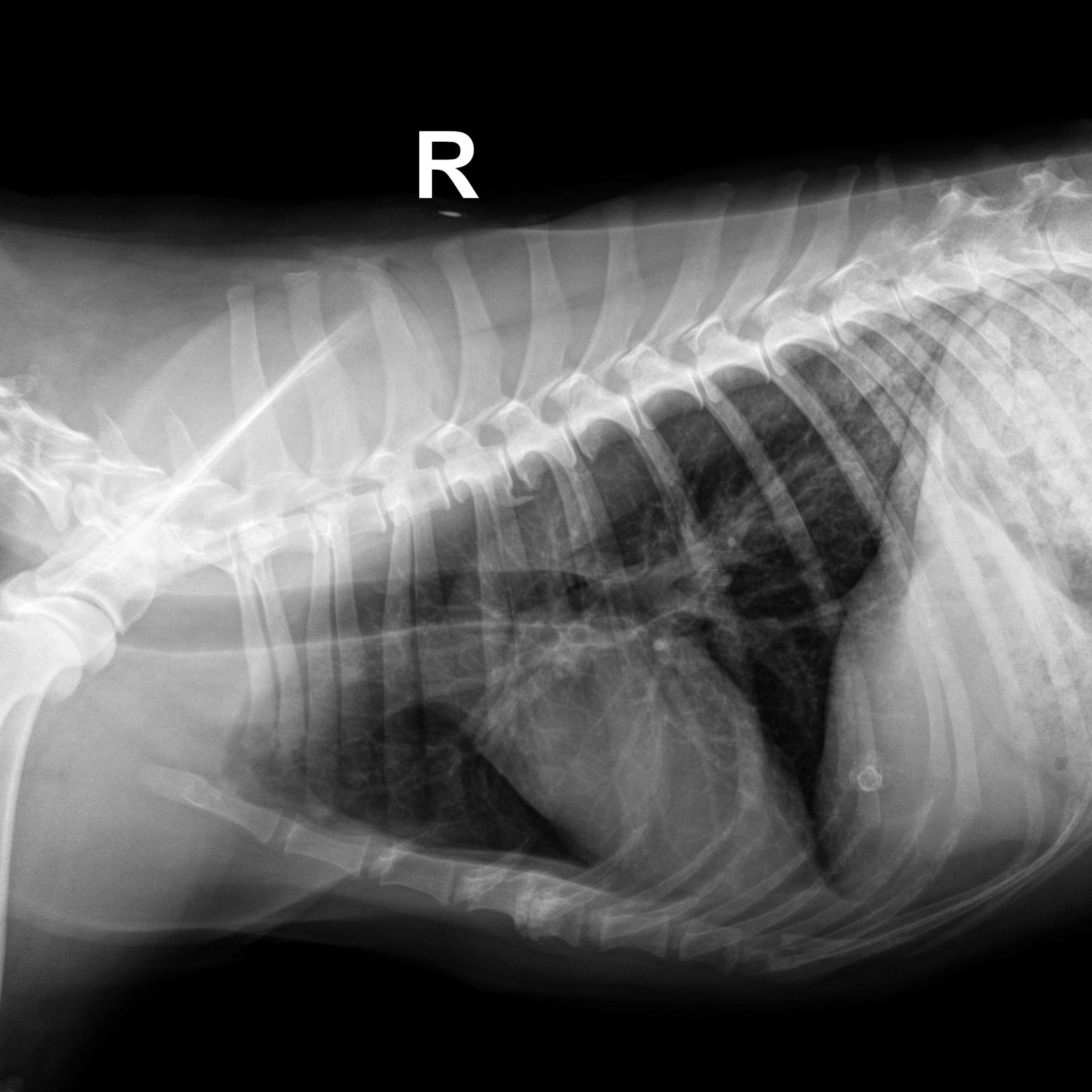
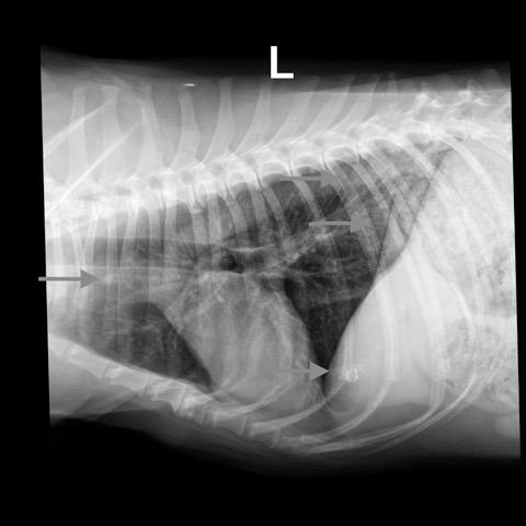
The findings indicate mild non-cardiogenic (consider neurogenic) pulmonary edema. There was hypovolemia with microcardia and underperfusion of the lung and a chronic bronchial pattern, which may be age related or associated with chronic airway disease. The small nodule is compatible with an end on pulmonary vessel. A secondary neoplastic nodule cannot be ruled out. The mineral opaque material is likely associated with the hepatic biliary system. This is usually benign. Correlate with the chem profile.

Image Interpretation

Rads of right lateral, left lateral and VD thorax – Osseous structures: There was mild ventral vertebral endplate modeling within the
mid thoracic spine consistent with spondylosis deformans.
Extrathoracic soft tissue structures: There was a spherical mineral opaque structure
and granulated mineral opaque material superimposed onto the cranial aspect of the
liver and caudal aspect of the lung respectively.
Intrathoracic structures: The course of the trachea was normal. The cardiac silhouette
was small, the caudal vena cava and the pulmonary vasculature were thin.
There was an area of diffusely increased interstitial opacity within the caudodorsal
aspect of the lung.
There also was a generalized bronchial pattern with increased wall visibility and
mineralization.
There was a small soft tissue opaque nodule associated with the cranial lobar vessels
best visible on the left lateral view (see image below)
Comments

A total number of 3 views were available for assessment in jpeg format – DICOM would be the preferred format for unlimited postprocessing. The degree of inspiration was moderate. Moderate motion artifact and low signal to noise ratio due to underexposure on the VD view.

Patient Information

Patient Name : Mushka Webber, Critical Vet Care
Gender : Male, Neutered
Species : Canine
Type of Imaging : Ultrasound
Status : Complete

Clinical Signs

  • Seizures

Images

a0500000094475_ma0500000094476_ma0500000094476_ma0500000094477_m

Blood Chemistry

  • Glucose, High

Clinical Signs

  • Seizures
Skip to content