RADS – Aggressive Osteolytic Ulnar Bone Lesion in a 13 year old FS Samoyed dog

Case Study

RADS – Aggressive Osteolytic Ulnar Bone Lesion in a 13 year old FS Samoyed dog

Presenting Clinical Signs: 2 month history of intermittent left forelimb lameness progressing to non-weight bearing lameness left forelimb past 2 days.

Physical Exam: Severe discomfort on flexion of left elbow, firm mammary mass (owner declines work up for suspected mammary tumor- patient was spayed 5 years ago) . CBC, Chem, UA pending
Presenting Clinical Signs: 2 month history of intermittent left forelimb lameness progressing to non-weight bearing lameness left forelimb past 2 days.

Physical Exam: Severe discomfort on flexion of left elbow, firm mammary mass (owner declines work up for suspected mammary tumor- patient was spayed 5 years ago) . CBC, Chem, UA pending

Image Interpretation

mediolateral left front limb, mediolateral and craniocaudal view right front limb – Left front limb:
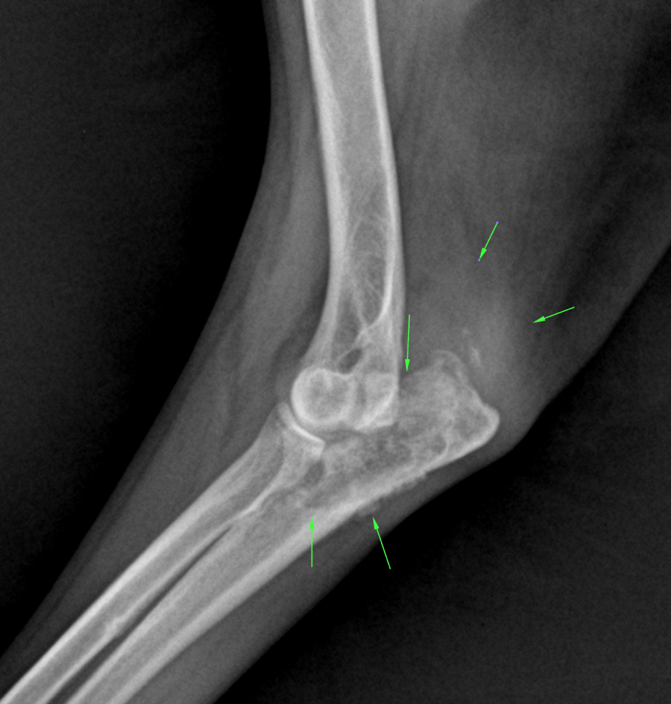
There was a soft tissue swelling with amorphous new bone formation localized to the area proximal to the olecranon. The delineation of the triceps muscle insertion was indistinct. An area of permeative osteolysis with endosteal scalloping, multifocal cortical thinning, and amorphous periosteal reactions was associated with the proximal ulna epiphysis and metaphysis. The transition zone to the healthy bone was long and indistinct. Significant muscle atrophy was noted in the left front limb.
A smooth osteophyte was present at the caudal contour of the humeral head consistent with mild osteoarthrosis. The intertubercular groove presented a focal increase in bone opacity suggesting chronic biceps tendinopathy. The carpal joint presented mild osteoarthrotic changes.
Right front limb: The carpal joint presented mild osteoarthrotic changes.

DX

The radiographic findings are compatible with a monostotic aggressive osteolytic lesion within the proximal ulna. Primary and secondary neoplasia of bone has to be considered as underlying cause.

Comments

Secondary neoplasia, such as metastatic spread of another primary tumor, is by far more likely owing to the unusual location for a primary bone neoplasm and the history of the mammary neoplasia. Differentials for a potential primary neoplasia of bone would include osteosarcoma, fibrosarcoma, hemangiosarcoma, chondrosarcoma. In case the dog was in an endemic region fungal osteomyelitis would be the only potential differential diagnosis to a orimary or secondary malignancy of the bone. Arthritis can be ruled out as an underlying cause based on the radiographic findings. 

Apart from the further evaluation of the mammary neoplasia a full tumor staging would have to include chest radiographs, abdominal ultrasound, regional lymph node evaluation and tumor sampling. 

Please note the left ulna is prone to pathologic fracture! 

Patient Information

Patient Name : Jubilee Rampersand, Perry Hall AH
Gender : Female, Spayed
Species : Canine
Type of Imaging : Ultrasound
Status : Complete
Liz Wuz Here : Yes

Clinical Signs

  • Lameness

Exam Finding

  • Masses

Images

bildschirmfoto_2015-09-16_um_12

Clinical Signs

  • Lameness
Skip to content