This 4 year old MN Dalmation dog presented with spontaneous acute respiratory distress, foaming at mouth, short shallow breathes. Decompensated within 10 min and crashed.
Noted the pneumothorax. Was unable to get DV or VD fast enough.
Test tapped for air but with this patient just wasn’t quick enough. No punctures noted. Have you ever seen a spontaneous pneumothorax like this before?
No known sharp object ingestion.
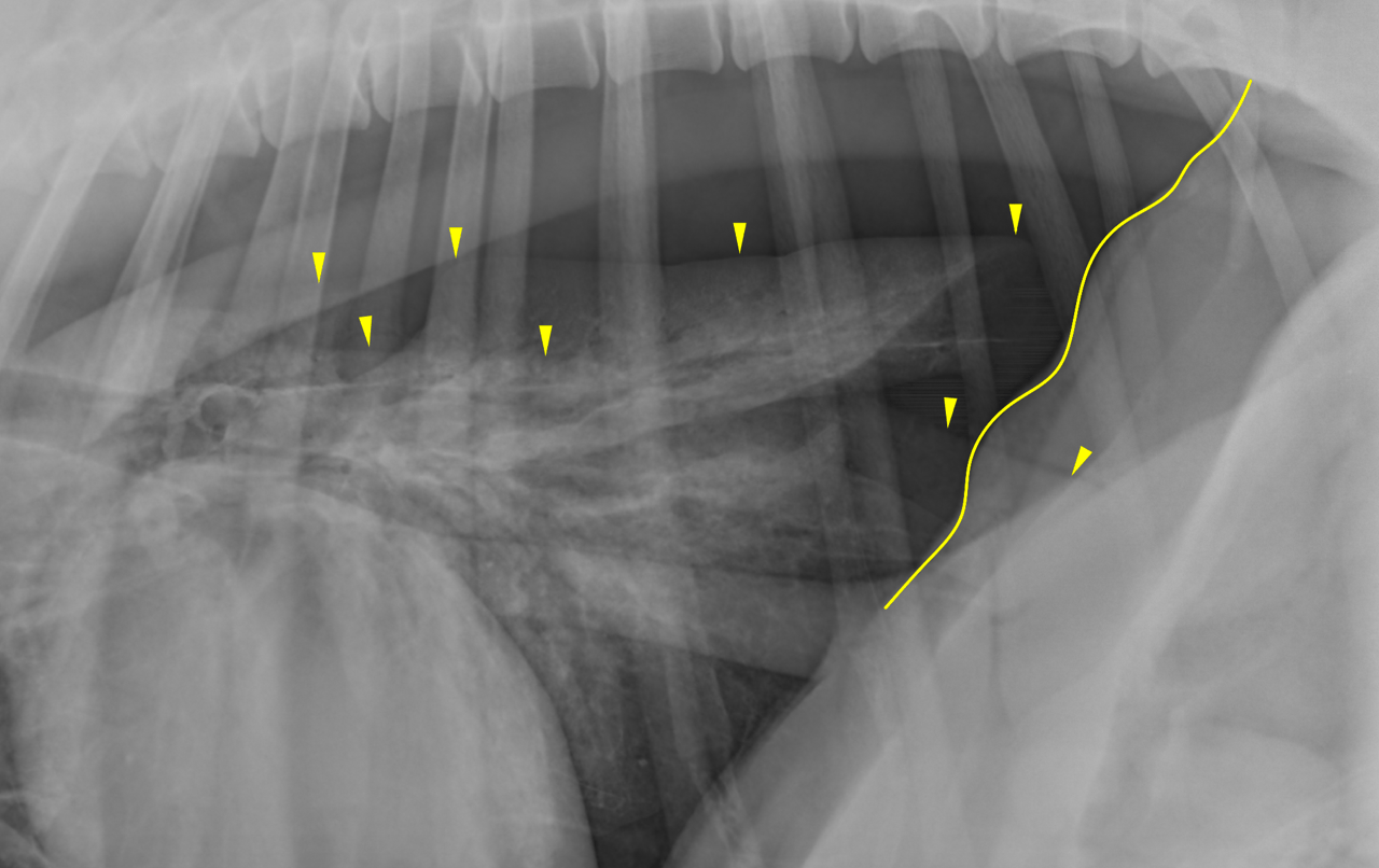