RAD – Normal juvenile thorax in a 6 month old Golden Retriever dog with tall R waves on ECG

Case Study

RAD – Normal juvenile thorax in a 6 month old Golden Retriever dog with tall R waves on ECG

This 6 month old Golden Retreiver puppy was presented for an ovariohysterectomy. Tall R waves were noted on an ECG. Physical exam wnl. Need confirmation of no cardiac abnormalities prior to spay.
This 6 month old Golden Retreiver puppy was presented for an ovariohysterectomy. Tall R waves were noted on an ECG. Physical exam wnl. Need confirmation of no cardiac abnormalities prior to spay.

Image Interpretation

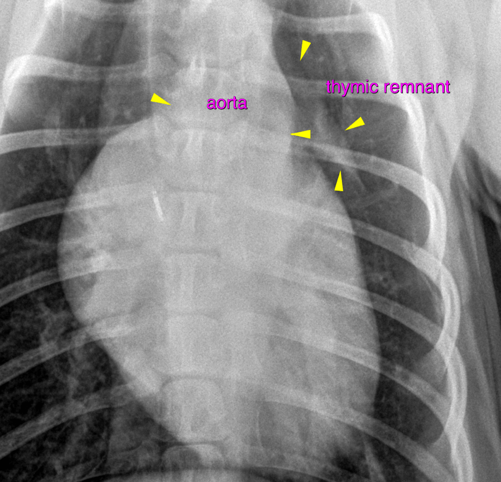
Rads – Lateral and VD thorax – Soft tissue opaque cranial mediastinal widening with a veil shaped structure border effacing with the heart is noted and compatible with a thymic remnant. Even though the pulmonary vasculature of the left caudal lobe seems prominent on the ventrodrosal view, the overall impression of the pulmnoray vasculature and perfusion is within normal limits. A mild amount of air is seen within the non-dilated esophagus cranial to the heart which is within normal limits.

DX

Normal juvenile thorax

Outcome

There is no radiographic evidence of cardiac disease. The list of possible differential diagnoses for tall ECG R-waves includes diseases that cannot be assessed by means of radiographs alone – such as ventricular hypertrophy in subaortic stenosis or other congenital anomaly, muscular dystrophy, and more. Comprehensive screening for hemodynamic changes by means of a full cardiac echo is strongly advised in case the heart auscultation is abnormal

Patient Information

Patient Name : Harper Bischoff/BCVC
Gender : Female, Intact
Species : Canine
Type of Imaging : Ultrasound
Status : Complete
Liz Wuz Here : Yes
Code : 17_00003

Images

bildschirmfoto_2016-05-04_um_15
Skip to content