This 5 year old MN ferret presented with an elevated respiratory rate, grade 2/6 murmur, sub lumbar mass, enlarged heart
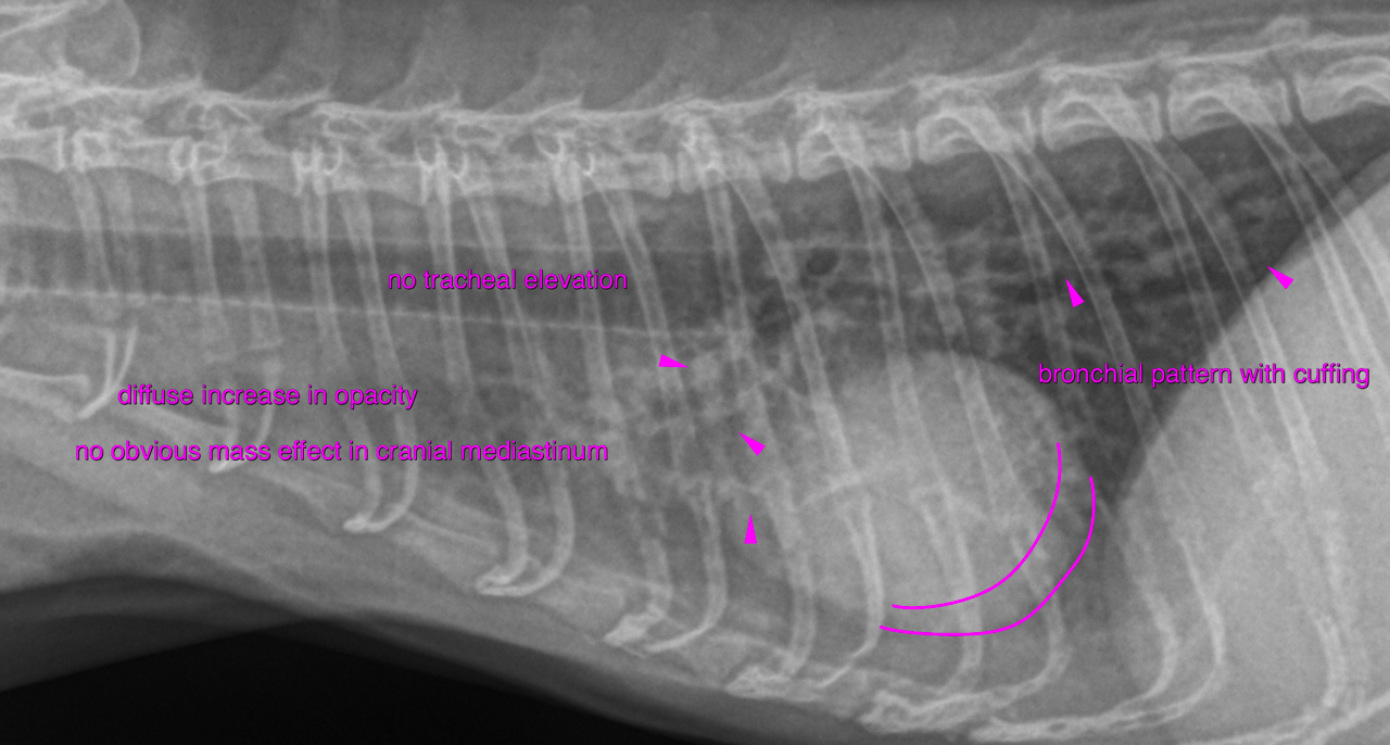
Rads of the thorax – A moderate multifocal bronchial pattern with peribronchial cuffing is noted.
The cranial mediastinum reveals widening at 2 times the width of the spine. The
cranial mediastinum and pericardium reveal double opacity contours indicative of
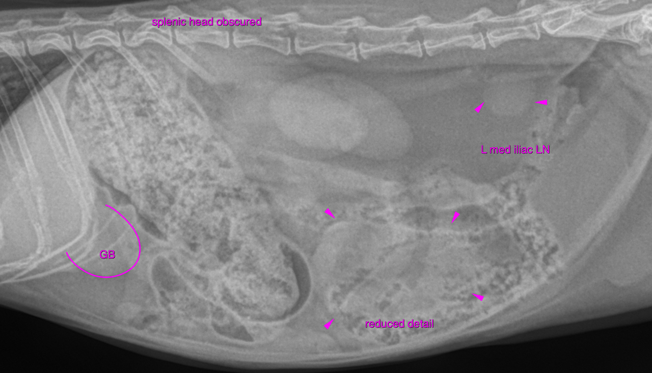
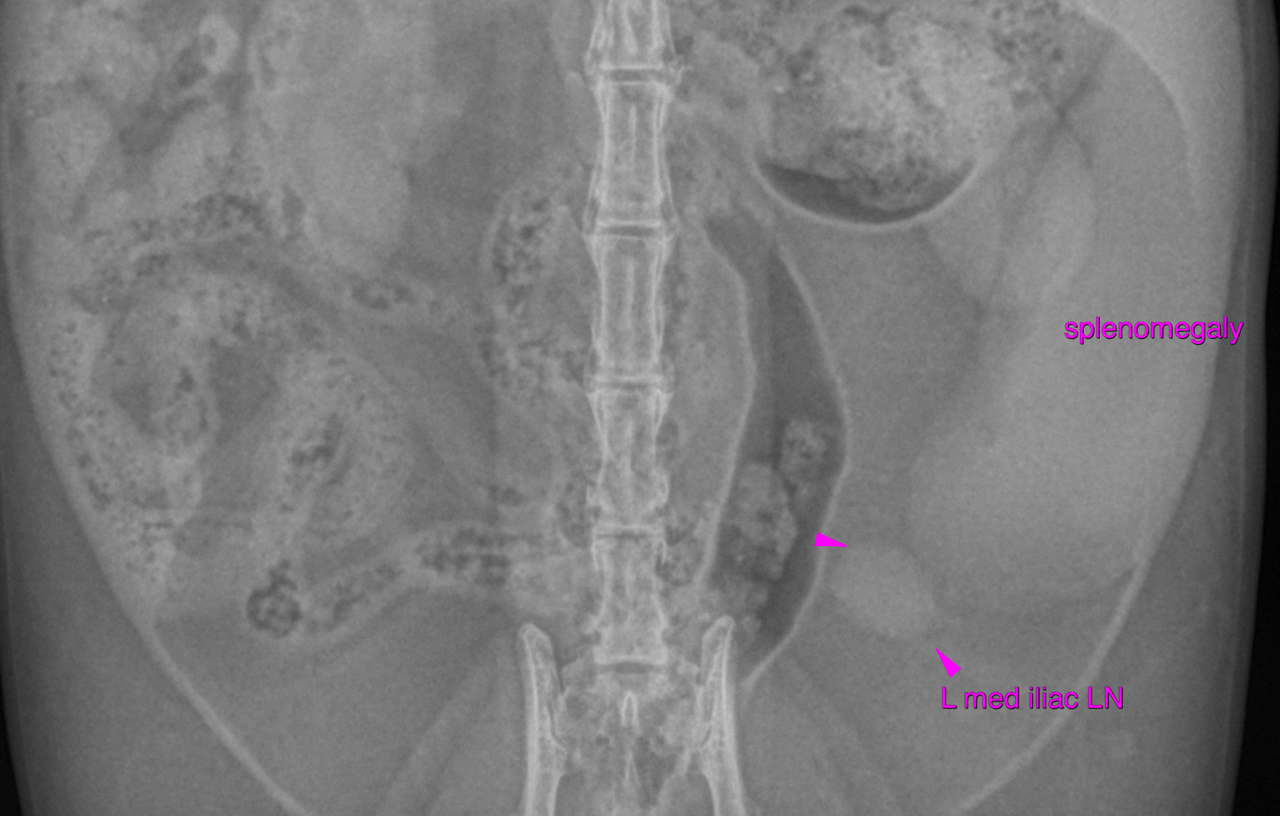
mediastinal fat. There is moderate generalized splenomegaly with rounded margins. The splenic head
is obscured on the lateral view, which is likely owing to the mass effect.
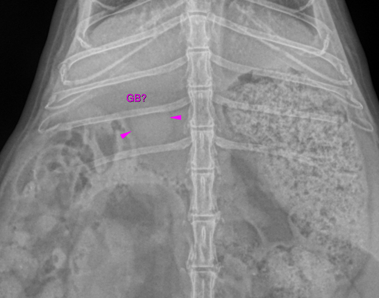
A round soft tissue opaque structure is seen between the liver and the moderately
distended and ingesta filled stomach, which is likely to be compatible with the
distended gallbladder. Theoretically this could represent a cranial abdominal
lymphadenomegaly or cyst as well. The left medial iliac lymph node reveals moderate enlargement and rounding.
The findings are suggestive for a multifocal neoplastic infiltrate involving the spleen
and lymph nodes. The bronchitis may be infectious (bacterial, viral, parasitic) or allergic in origin.
Testing for lungworms is advised. Consider further workup with tracheal wash or
bronchoalveolar lavage if the patient is refractory to symptomatic treatment.