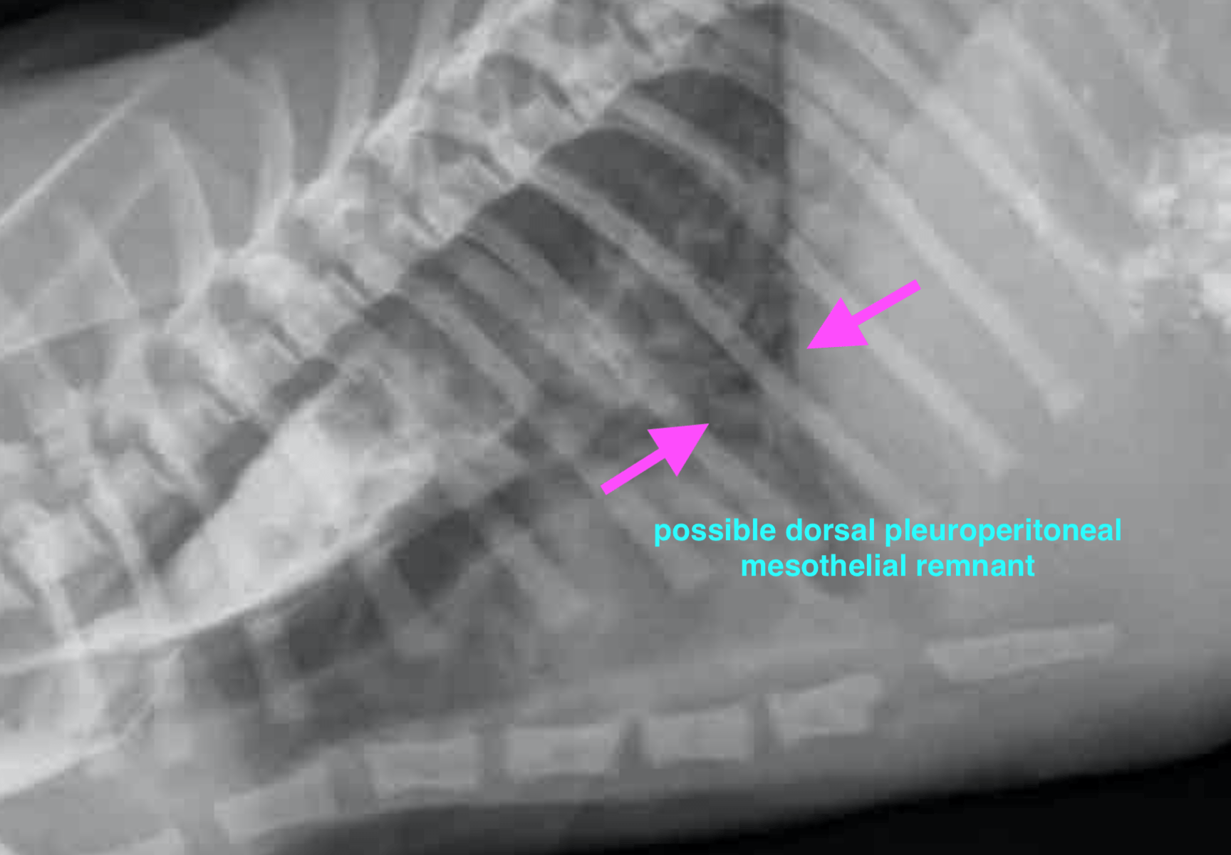
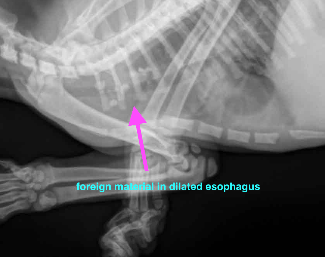
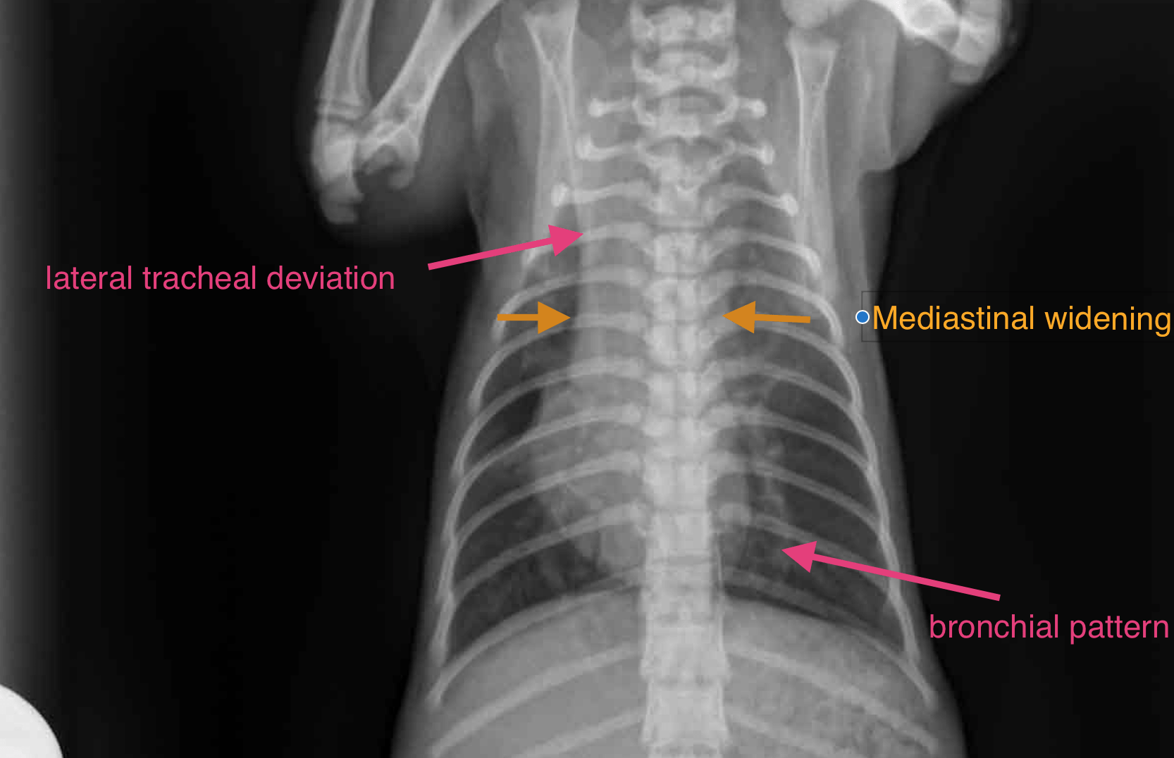
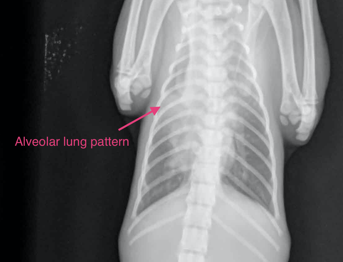
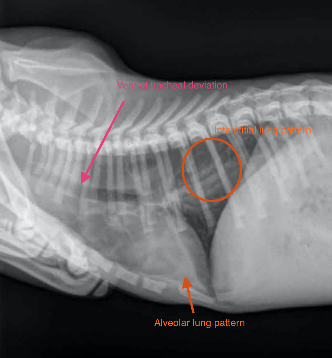
This 5 month old M kitten has a history of chronic vomiting of solid food, can only keep down liquids. Has been extensively dewormed. Felv/fiv neg, TP 5.1 low, albumin 2.4 low, globulin 2.2 low, neutrophilia and eosinophilia.
This 5 month old M kitten has a history of chronic vomiting of solid food, can only keep down liquids. Has been extensively dewormed. Felv/fiv neg, TP 5.1 low, albumin 2.4 low, globulin 2.2 low, neutrophilia and eosinophilia.