Rads- lateral and VD thorax, lateral abdomen -Osseous structures:
Overall moderate degenerative changes were associated with the axial skeleton including narrow disc spaces with endplate spondyloses at the cervicothoracic transition and mid thoracic spine. Mild osteoarthritic changes were seen at the shoulder joints.
Both shoulder joints and elbow joints revealed moderate and mild osteoarthritic changes respectively. The chest wall revealed focal thickening in the region of the right armpit which likely was due to the positioning of the right front limb.
Intrathoracic structures:
The views were obtained during expiration.
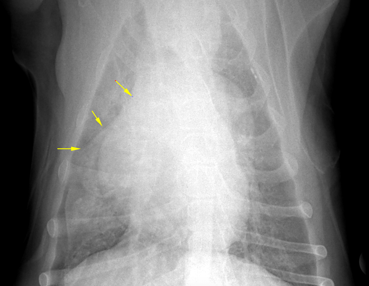
The trachea, mediastinum and pleural space were within normal limits. The lungs revealed a moderate generalized increase in interstitial opacity emphasizing the right caudal and the right middle lobe. The heart was mildly shifted towards the right side owing to volume loss of the right middle lobe. Multifocal bronchial wall mineralization was noted. No interstitial nodular changes were seen.
The cardiac silhouette was within normal limits for size and shape. The major vessels and pulmonary vasculature were within normal limits. There was no sign of vascular congestion.
Intraabdominal structures:
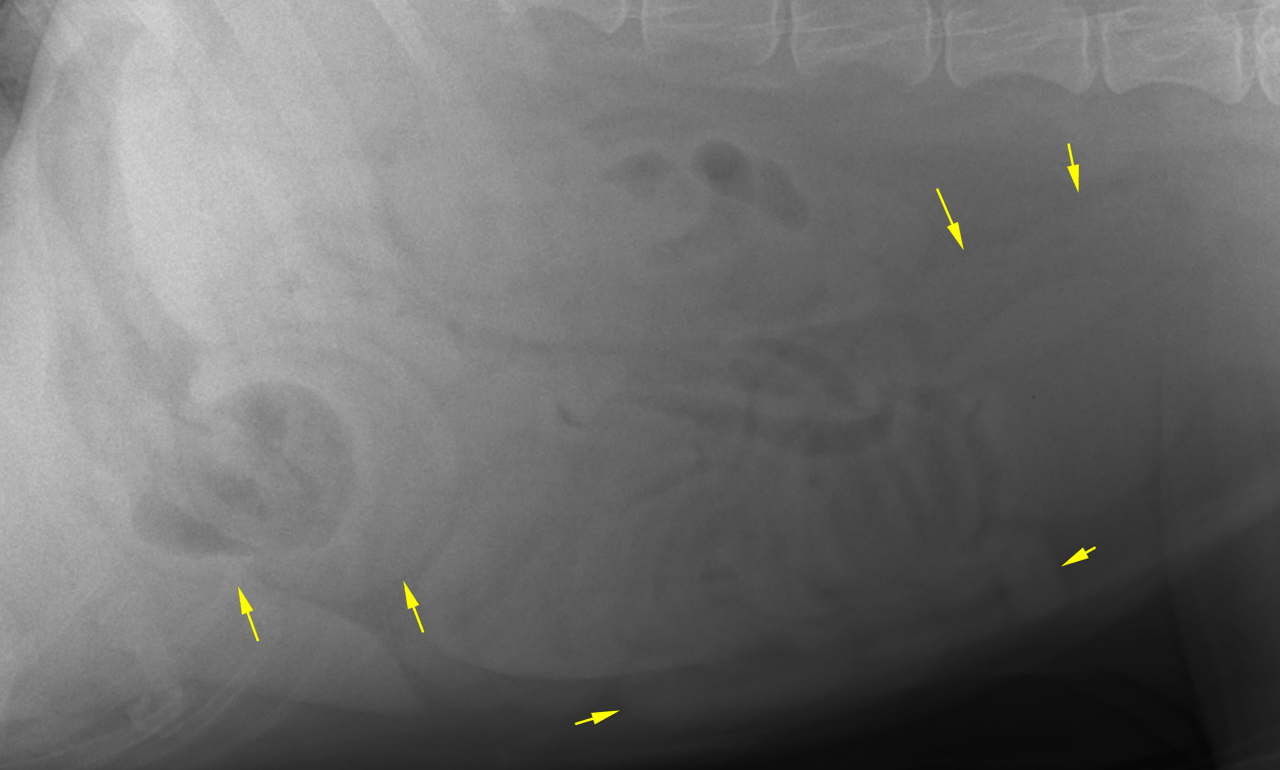
The serosal detail was within normal limits.
The liver extended beyond the costal arch, but the liver margins were pointed.
The kidneys, prostate and urinary bladder were within normal limits.
The stomach was contracted and contained a mild amount of gas and fluid. The small intestinal loops were non dilated and of even diameter but presented a maldigestion pattern.
The colon was largely empty.