RAD – Cranial cruciate repair failure with possible meniscal damage in a 6 year old MN American Staffordshire Terrier

Case Study

RAD – Cranial cruciate repair failure with possible meniscal damage in a 6 year old MN American Staffordshire Terrier

This 6 year old American Staffordshire Terrier has a history of left CCL repair from 2014. Presently non-weight bearing left hind with quadriceps contracture. R/O meniscal tear

This 6 year old American Staffordshire Terrier has a history of left CCL repair from 2014. Presently non-weight bearing left hind with quadriceps contracture. R/O meniscal tear

Image Interpretation

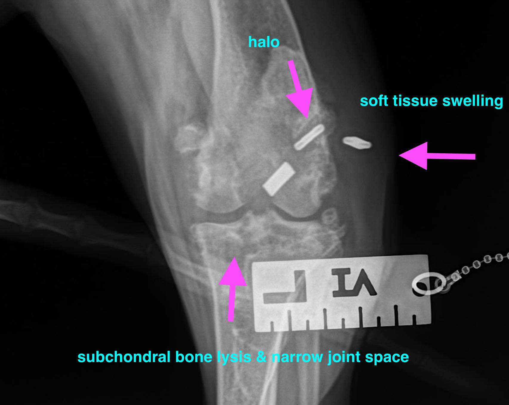
The left stifle joint presents a status after surgical repair of a cranial cruciate ligament pathology. 3 metal opaque devices are seen, 2 of them associated with a drill hole within the lateral femoral condyle, one of them within the soft tissue lateral to the stifle joint. The implants within the lateral femoral condyle are surrounded by a thin radiolucent halo. The device within the soft tissue is surrounded by a local soft tissue swelling and an Überschwinger artifact.

A mild articular soft tissue swelling is seen. The periarticular margins present a large amount of new bone formations. Multifocal geographic osteolysis is seen within the subchondral bone of the femorotibial joint. The femorotibial joint space presents narrow. Faint mineralization is seen  associated with the meniscal area. The left thigh musculature as well as the left femur and tibia present severe generalized disuse atrophy.

A mild articular soft tissue swelling is seen associated with the right stifle joint. A moderate amount of osteophytes is present at the periarticular margins. Multifocal mineralization and emerging meniscal mineralization is seen associated with the right stifle joint.
DX

left stifle- • Status after surgical repair of cranial cruciate ligament failure with advanced degenerative joint disease and chronic activated/intermittent arthritis • Indirect evidence of meniscal damage and/or compartment syndrome • Severe disuse muscle and bone atrophy • Possible implant failure with soft tissue reaction/inflammation

Outcome

Radiographically in the left stifle there is no strong indication of septic arthritis/cellulitis. However, complementary aspiration of synovia for cytology and culture is recommended as well as clinical evaluation for sifle joint instability. Meniscal damage appears likely. The right stifle shows chronic degenerative joint disease and indirect evidence of meniscal degeneration

Patient Information

Patient Name : Wrigley/Wauwatosa
Gender : Male, Neutered
Species : Canine
Type of Imaging : Ultrasound
Status : Complete
Liz Wuz Here : Yes
Code : 17_00057

Clinical Signs

  • Lameness

Images

bildschirmfoto_2016-08-12_um_15

Clinical Signs

  • Lameness