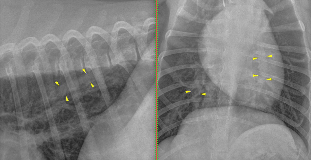
The patient is an 18 month old FS mixed breed dog that is heartworm positive. Has been on ivermectin (Heartgard plus) every 2 weeks for the last 8 months. Rads today to stage heartworm disease for consideration of immiticide treatment.
The patient is an 18 month old FS mixed breed dog that is heartworm positive. Has been on ivermectin (Heartgard plus) every 2 weeks for the last 8 months. Rads today to stage heartworm disease for consideration of immiticide treatment.