History: coughing for 2 weeks
History: coughing for 2 weeks
History: coughing for 2 weeks
History: coughing for 2 weeks
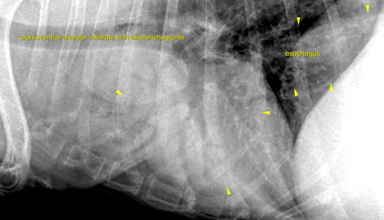
Rads of the thorax – There is a severe bilateral alveolar consolidation of the cranioventral and perihilar lung
field highlighted by airbronchograms, which mainly involves the left and right cranial
as well as the right middle lung lobe. Only the perihilar region of the caudal lung lobes
reveals an increased opacity, the remainder of the caudal lung field reveals the
expected age related changes.
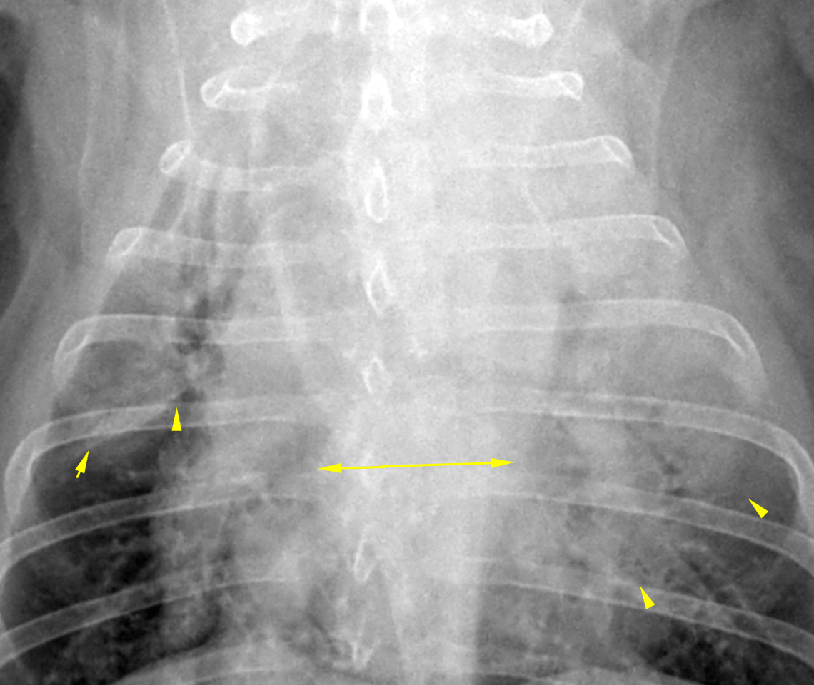
There is evidence of tracheobronchial lymph node enlargement indicated by split of the
main stem bronchi seen on the ventrodorsal view.
The cardiovascular structures are normal with no evidence of cardiomegaly, overt
chamber enlargement or congestive heart failure.
The cranial thoracic esophagus contains a mild amount of gas. The caudal thoracic
esophagus is seen as a mildly dilated soft tissue opacity, which likely is an incidental
finding associated with swallowing or gastroesophageal reflux during exposure.
The included bony and abdominal structures are within normal limits.
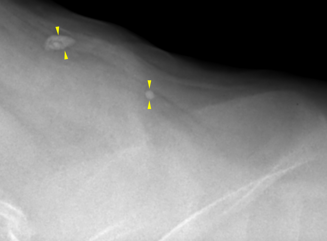
Two amorphous ovoid subcutaneous calcifications are seen dorsal to the scapulae with
no surrounding soft tissue swelling. This is likely consistent with incidental dystrophic
mineralization or calcinosis cutis.
Severe bilateral alveolar consolidation of the cranioventral lung field with suspected
mediastinal lymph node enlargement.
The findings are suggestive of infectious pneumonia. Bacterial, mycobacterial and
fungal microorganisms need to be considered.
The main differential diagnosis is a secondary neoplasia of the lung such as round cell
neoplasia: lymphoma or histiocytic sarcoma.
Pulmonary hemorrhage and aspiration pneumonia do not match the symmetrical
distribution of the infiltrate.
Bronchoscopy with bronchoalveolar lavage plus ultrasound guided fine needle
aspiration of the lung is recommended for further definition. In case the owner declines
bronchoscopy/sedation ultrasound guided fine needle aspiration of the lung followed
by symptomatic treatment is recommended as a diagnostic challenge. Check
coagulation profile.