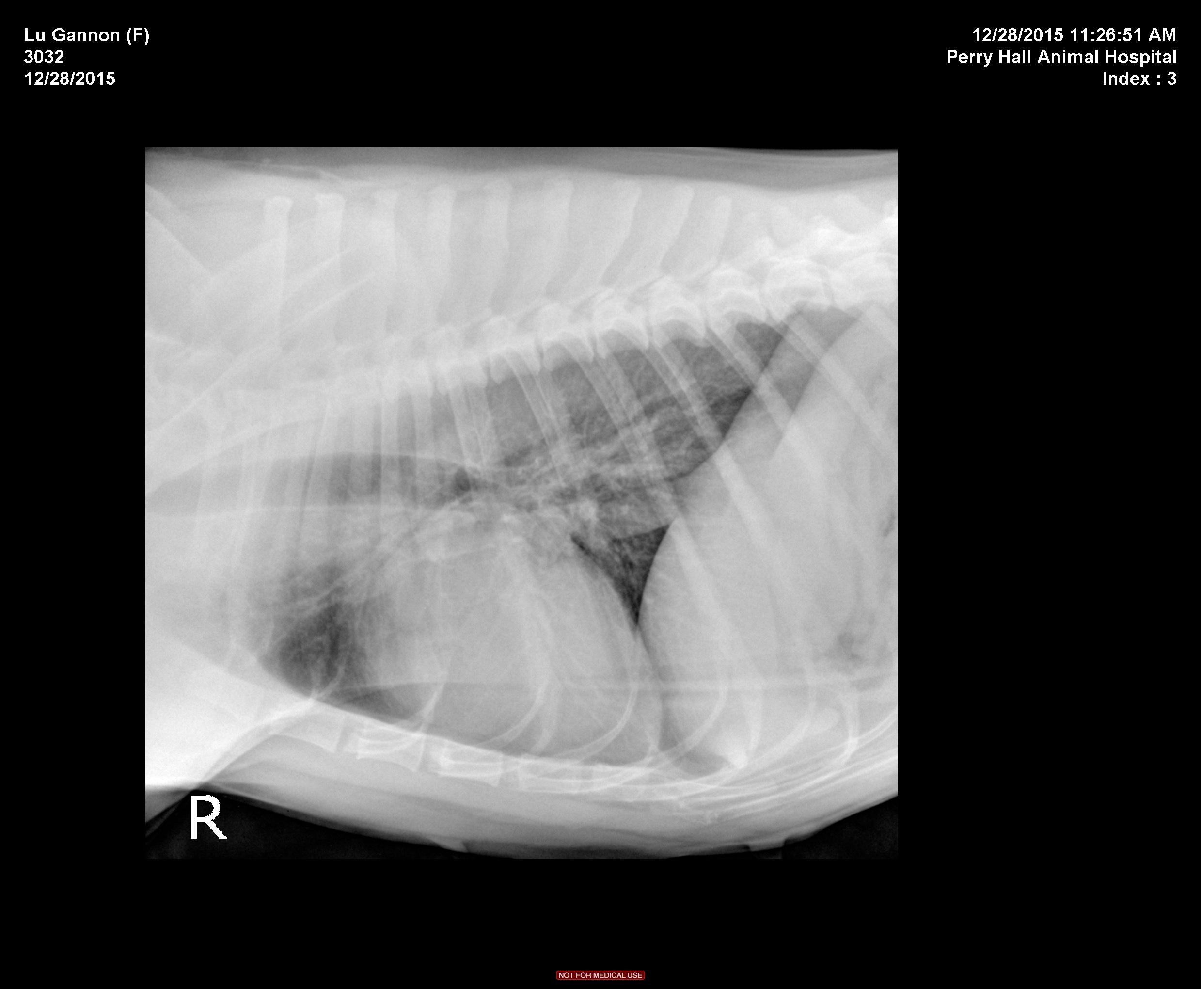
The patient is a 9 year old FS Pitbull with a 3 day history of lethargy and decreased appetite. Has not eaten in 24 hours, decreased water intake. Seems almost ataxic or weak to the owner at home, overall very depressed. Hx of allergies that are controlled.
Physical Exam: Depressed, Temp 101.9. MM pink/slightly tacky. cardiac wnl, lungs wnl on auscultation. BCS 5/9. No abdominal discomfort on palpation. Walking normally x 4 in room but is much less energetic than usual.
CBC/chem pending
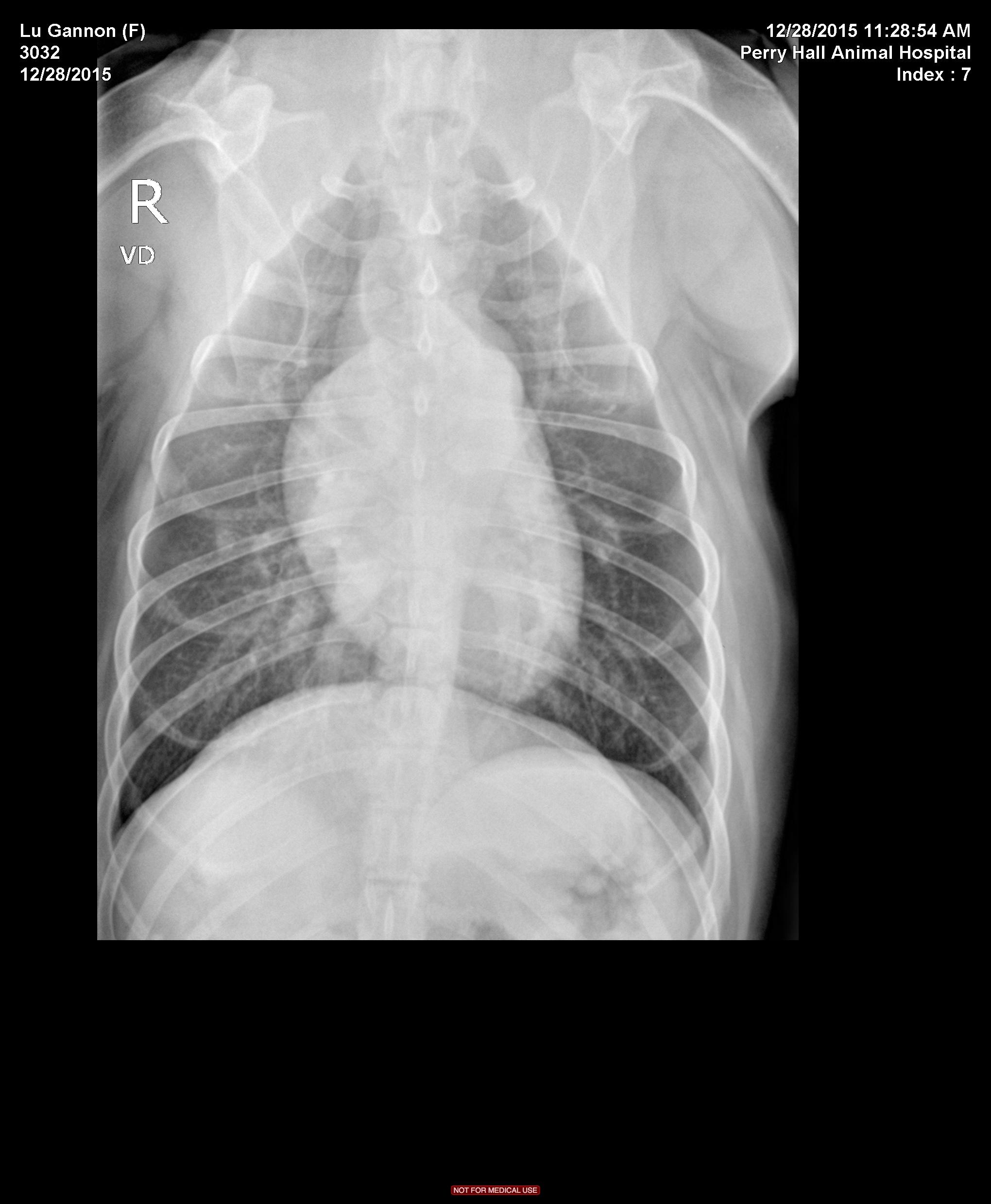