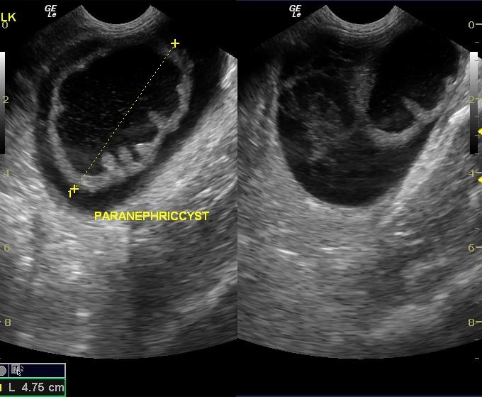
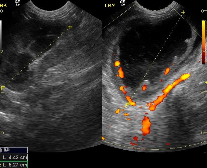
A 5-year-old FS DLH cat was presented for hiding at home and anorexia. Abnormalities on physical examination included a large urinary bladder and mild dehydration. Leukocytosis was present on CBC; blood chemistry was within normal limits. Survey abdominal radiographs showed left-sided renomegaly and a small right kidney.
A 5-year-old FS DLH cat was presented for hiding at home and anorexia. Abnormalities on physical examination included a large urinary bladder and mild dehydration. Leukocytosis was present on CBC; blood chemistry was within normal limits. Survey abdominal radiographs showed left-sided renomegaly and a small right kidney.