An 11-year-old FS Yorkshire Terrier dog, currently on hydroxyzine, was presented for polypnea while at rest. On physical examination a grade III/VI systolic murmur and clear lungs fields were present.
An 11-year-old FS Yorkshire Terrier dog, currently on hydroxyzine, was presented for polypnea while at rest. On physical examination a grade III/VI systolic murmur and clear lungs fields were present.
Case Study
Pulmonary hypertension (Severe) & aortic body tumor in a 11 year old FS Yorkshire Terrier dog
Sonographic Differential Diagnosis
Severe pulmonary hypertension, aortic body tumor.
Image Interpretation
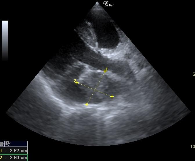
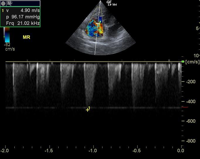
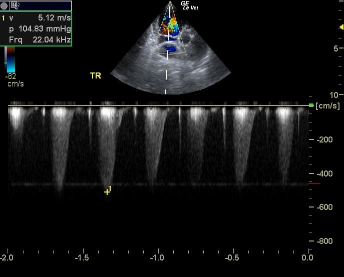
The echocardiogram presented a prominent right heart with mild right ventricular hypertrophy without clinically significant tricuspid regurgitation, and normal right atrial size. Severe tricuspid insufficiency was noted at 5.12m/sec. This is consistent with severe pulmonary hypertension. The patient is at high risk for sudden death. No evidence of neoplasia was noted in the right auricle, or elsewhere in the heart. The pulmonary artery was uniformly prominent with mildly depressed pulmonic velocity measured on PW Doppler. The mitral valve presented vegetative contour and was visibly insufficient on PW and color flow Doppler. Severe mitral insufficiency was noted at 4.9 m/sec. The left atrial size was enlarged and there an aortic body tumor that measured 2.8 cm. This impinged into the left atrium. The left ventricular outflow demonstrated normal flow patterns and velocities through the aortic valve.
DX
Severe pulmonary hypertension, aortic body tumor.
Outcome
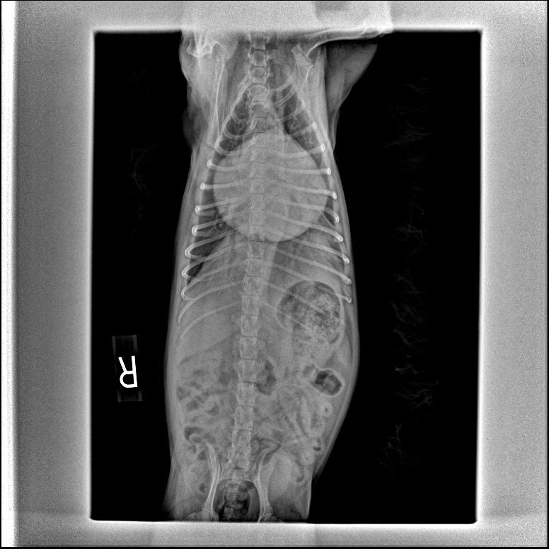
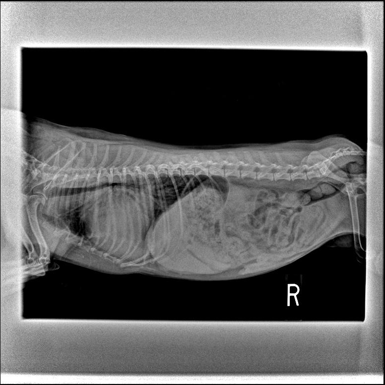
Thoracic survey radiographs showed a severely enlarged cardiac silhouette and perihilar edema. Systemic hypertension was present on blood pressure measurement (270/155). The patient was treated with Enacard, Lasix, and Viagra. A very guarded long term prognosis was given and the owners were warned about the risk of sudden death in this patient. At a recheck appointment a week later the patient`s blood pressure was 155/80 and a slight wheeze was asculted. Thoracic radiographs showed ongoing edema and an enlarged cardiac silhouette.
Clinical Differential Diagnosis
Pulmonary edema secondary to heart disease, pleural effusion, pulmonary embolism, primary lung disease – pneumonia/fibrosis/allergic disease
Video
Patient Information
Patient Name :
Fiona O
Gender :
Female, Spayed
Species :
Canine
Type of Imaging : Ultrasound
Status :
For Review
Liz Wuz Here :
Yes
Code :
15-00003
Clinical Signs
- Tachypnea
Exam Finding
- Heart Murmur
Images
Clinical Signs
- Tachypnea