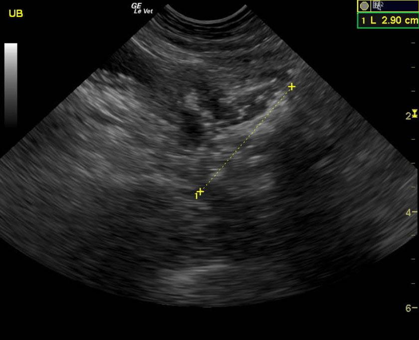
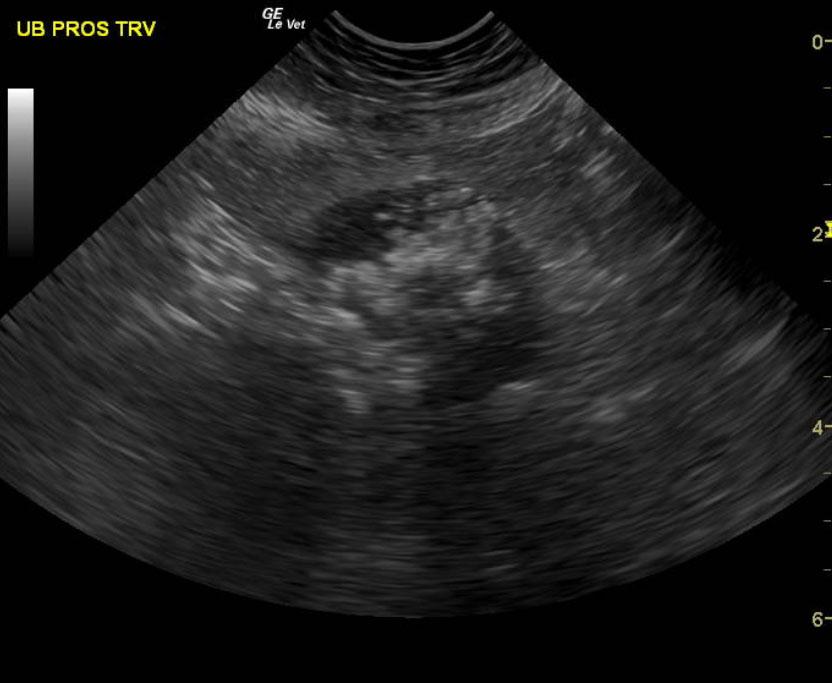
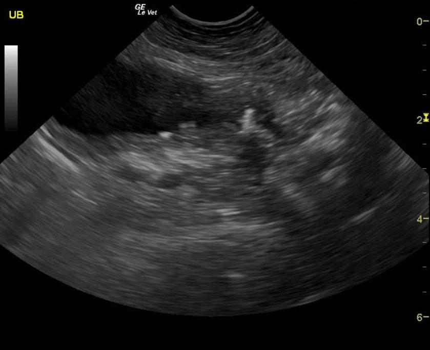
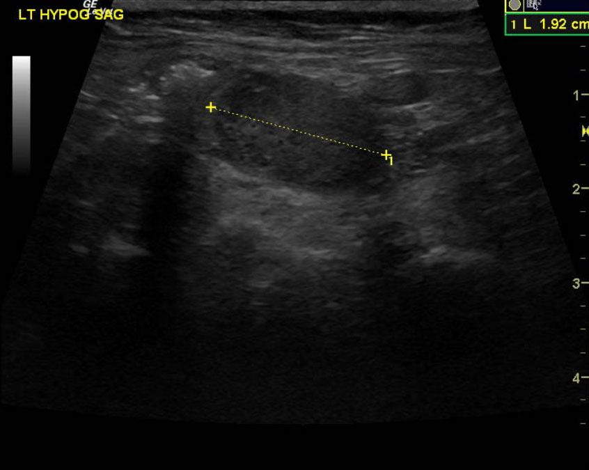
An 11-year-old neutered male Pomeranian dog was presented for evaluation of tenesmus, “hunched” stance, and decreased energy. On physical examination, a 3cm palpable mass cranial to pelvic canal was noted. Survey radiographs showed a mineralization/spherical mass in area of the prostate.
An 11-year-old neutered male Pomeranian dog was presented for evaluation of tenesmus, “hunched” stance, and decreased energy. On physical examination, a 3cm palpable mass cranial to pelvic canal was noted. Survey radiographs showed a mineralization/spherical mass in area of the prostate.