A 9-year-old MN DSH presented with anorexia and weight loss. Physical examination revealed poor body condition and mild dehydration. Moderately elevated SAP and mildly elevated ALT and mildly elevated AST were revealed on blood chemistry analysis.
A 9-year-old MN DSH presented with anorexia and weight loss. Physical examination revealed poor body condition and mild dehydration. Moderately elevated SAP and mildly elevated ALT and mildly elevated AST were revealed on blood chemistry analysis.
Case Study
Plasma cell neoplasia and hepatic lipidosis
Sonographic Differential Diagnosis
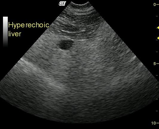
These findings are consistent with an infiltrative process. US-guided FNA revealed plasma cell neoplasia and moderate hepatic lipidosis.
Image Interpretation
Longitudinal view of the midliver, gallbladder (11 o’clock position), and falciform fat (top) reveals markedly diffuse hyperechoic parenchyma compared with the falciform fat.
DX
Plasma cell neoplasia and moderate hepatic lipidosis
Outcome
The patient was humanely euthanized owing to poor clinical response and owner reluctance to pursue chemotherapy.
Clinical Differential Diagnosis
Neoplasia, hepatitis, hepatic lipidosis, feline infectious peritonitis (FIP), pancreatitis, IBD.
Sampling
Owing to moderate PT and aPTT elevations, as well as the poor clinical status of the patient, US-guided FNA rather than biopsy was performed. The cytology revealed plasma cell neoplasia and moderate hepatic lipidosis.
Video
Patient Information
Patient Name :
Spanky E
Gender :
Male, Neutered
Species :
Feline
Type of Imaging : Ultrasound
Status :
Complete
Liz Wuz Here :
Yes
Code :
03_00006
Clinical Signs
- Anorexia
- Weight loss
Exam Finding
- Dehydration
- Weight loss
Images
Blood Chemistry
- Alkaline Phosphatase (SAP), High
- ALT (SGPT), High
- AST (SGOT), High
Clinical Signs
- Anorexia
- Weight loss