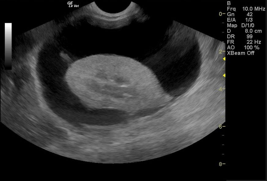
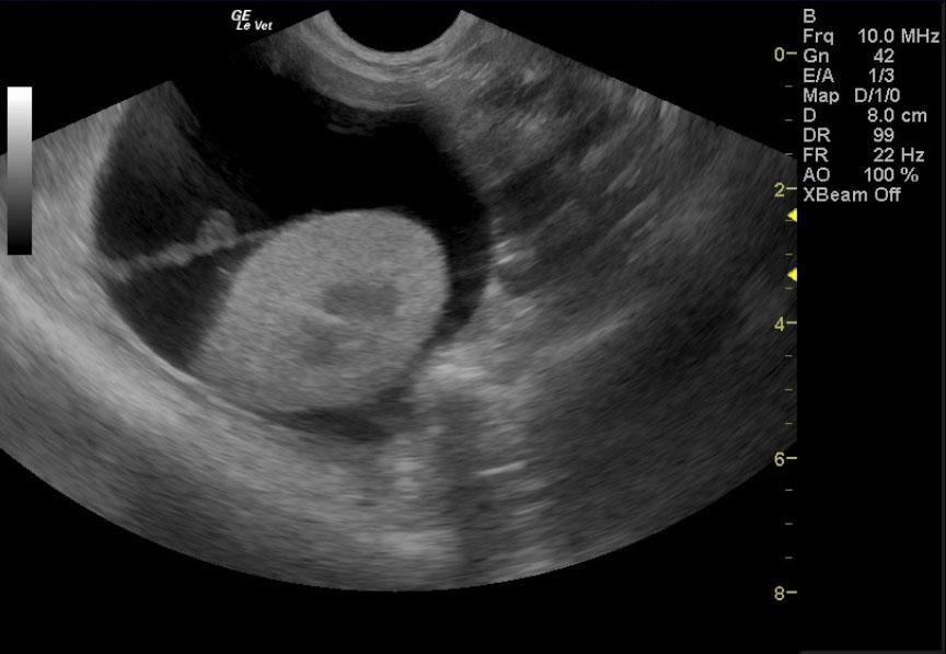
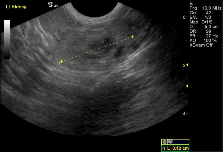
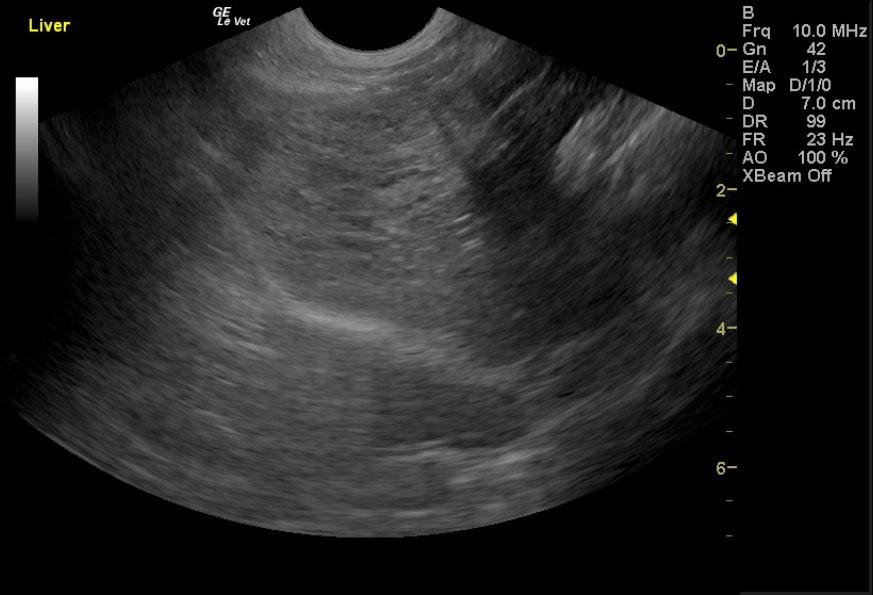
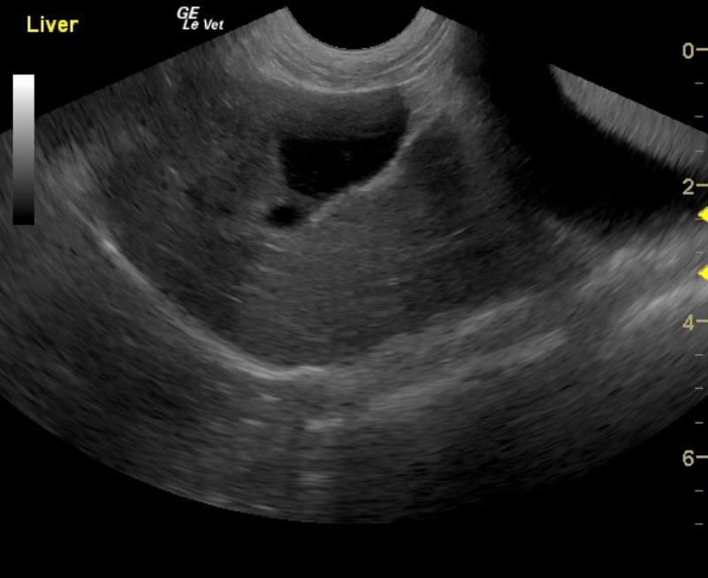
A 17-year-old neutered male DSH cat was presented to the emergency clinic with open-mouth breathing suspected to be secondary to stress/pain as the patient improved on analgesics. On a prior physical examination, an abdominal mass had been palpated.
A 17-year-old neutered male DSH cat was presented to the emergency clinic with open-mouth breathing suspected to be secondary to stress/pain as the patient improved on analgesics. On a prior physical examination, an abdominal mass had been palpated.