A 1-year-old intact female Yorkshire terrier was presented for evaluation of a grade V/VI left-sided heart murmur.
A 1-year-old intact female Yorkshire terrier was presented for evaluation of a grade V/VI left-sided heart murmur.
Case Study
Patent ductus arteriosis (PDA) and secondary pulmonic insufficiency in a 1 year old FI Yorkshire Terrier
Sonographic Differential Diagnosis
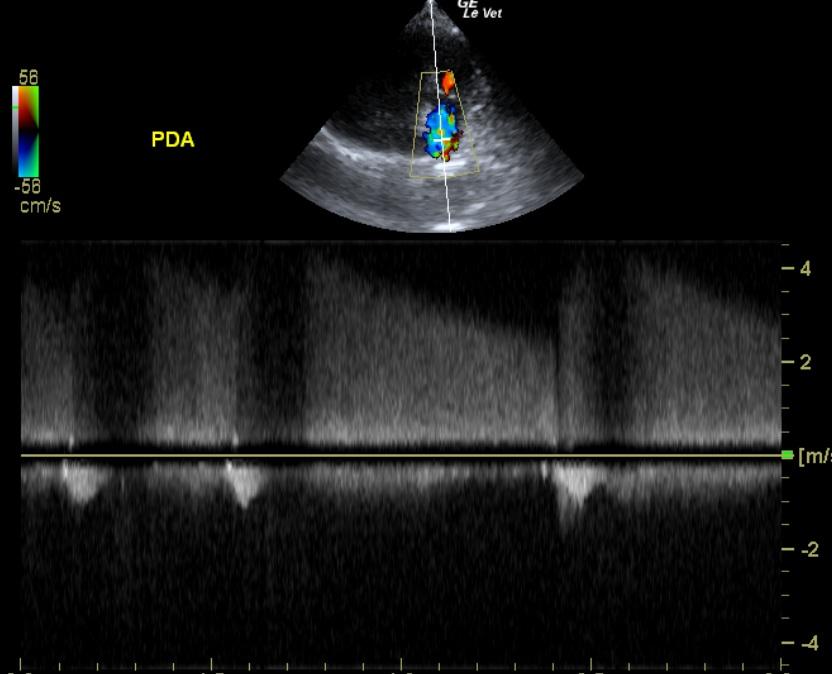
Patent ductus arteriosus. Secondary pulmonic insufficiency, which appears compensated at this time.
Image Interpretation
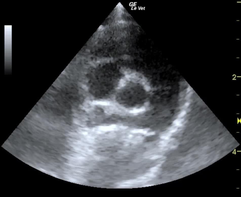
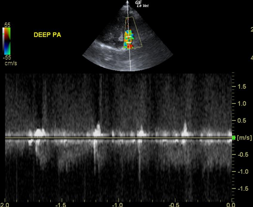
The right ventricle was of normal size (1/3 diameter of LV), echogenicity and thickness. There was no evidence of dilation or restriction. The right ventricular outflow tract was largely normal; however, the pulmonic valve, even though only slightly thickened, revealed significant insufficiency at 4.3 m/sec. Post-valvular forward flow was largely normal at 1.6 m/sec. However, the deep pulmonary artery (prior to the bifurcation) revealed a large amount of turbulence. Holosystolic flow was noted at 5 m/sec. This appears compensated at this time.
DX
PDA, secondary pulmonic insufficiency
Outcome
The owner was considering surgery to correct the PDA at the time of last communication.
Comments
A referral to a cardiologist would be recommended for potential coil placement or surgical intervention for PDA.
Clinical Differential Diagnosis
Congenital defect – PDA, atrial septal defect (ASD), ventricular septal defect (VSD), aortic stenosis, tricuspid dysplasia.
Sampling
None
Video
Patient Information
Patient Name :
Pixar H
Gender :
Female, Intact
Species :
Canine
Type of Imaging : Ultrasound
Status :
Complete
Liz Wuz Here :
Yes
Code :
15_00130
Clinical Signs
- Heart Murmur
Images
Clinical Signs
- Heart Murmur